Electrochemical investigation and surface engineering of living cells using ultramicroelectrodes require highly reliable probe positioning and accurate return-to-origin behavior. In probe-based systems such as Scanning Electrochemical Microscopy (SECM), SECM is a probing system used to measure chemical reactivity on the surface of the subject [1], small trajectory-dependent deviations in the positioning system can affect probe approach safety and measurement repeatability.
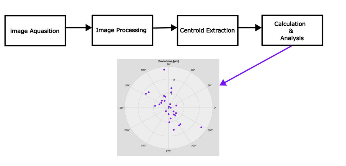
This study presents a vision-based approach for characterizing motion deviations in a Cartesian positioning system intended to support electrochemical probe-based experiments[2]. A CMOS camera was used to track the shadow of a platinum ultramicroelectrode at the return-to-origin position following repeated motion cycles. Image sequences were analyzed using a centroid-based intensity method to estimate positional deviations associated with different motion trajectories and feed rates.
The method was applied to representative linear and multi-axis trajectories relevant to probe positioning tasks. Experimental results show that, for most tested conditions, return-to-origin deviations remained below 2 µm, with maximum observed deviations on the order of 4 µm for certain trajectory and feed-rate combinations.
The results indicate that the vision-based deviation characterization enables reliable estimation of micrometer-scale positioning errors and provides a practical diagnostic tool for supporting ultramicroelectrode positioning in electrochemical investigations. This approach contributes to the development of stable motion platforms for living cell surface engineering and related probe-based micro-scale applications.