Electroporation (EP) is a promising technique for myocardial tissue modification, but its efficacy and safety depend on appropriate parameter selection, limited by the lack of practical methods for three-dimensional (3D) lesion assessment. This study aimed to establish a reliable framework for evaluating EP-induced myocardial lesions and identifying optimal EP parameters. Two complementary approaches were employed.
Microelectrode recordings at multiple sites within the EP-treated region determined transmural lesion depth by identifying where action potentials (AP) reappeared, and data from multiple experiments were integrated with AI-based analysis to reconstruct 3D lesion geometry. Lesion size was also assessed using optical mapping by analyzing optical action potential (OAP) amplitudes with depth-weighted exponential changes, and validated by ventricular wall sectioning and direct observation of AP propagation in the wedge.
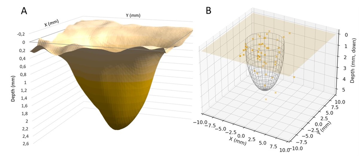
Using near-infrared di-4-ANBDQBS fluorescent dyes (depth constant around 2.1 mm) enabled signal detection and 3D assessment of lesion zones. Both methods enabled precise characterization of lesion dimensions, and optical mapping amplitude analysis (Figure 1A) reliably delineated lesion boundaries and closely matched microelectrode-derived transmural profiles (Figure 1B), providing faster and more reproducible evaluation.
These findings demonstrate that OAP amplitude measurements can be routinely applied to assess EP-induced lesion 3D zones and guide the optimization of EP parameters for effective and reproducible myocardial EP.