
Dynamic full-field optical coherence microscopy (d-FF-OCM) is a label-free imaging technique that allows to visualize cells and their intracellular activity in various fresh biomedical tissues. Images acquired by using these methods provide crucial information in various biophysical and biomedical research areas. However, d-FF-OCM systems do not come without their challenges when imaging biomedical, highly scattering tissues [1, 2]. In this work we present a high-resolution d-FF-OCM system (Fig. 1a) which overcomes most of the challenges and generates enhanced contrast images (Fig. 1b, c) without the need of any sort of fluorescence dyes [3]. 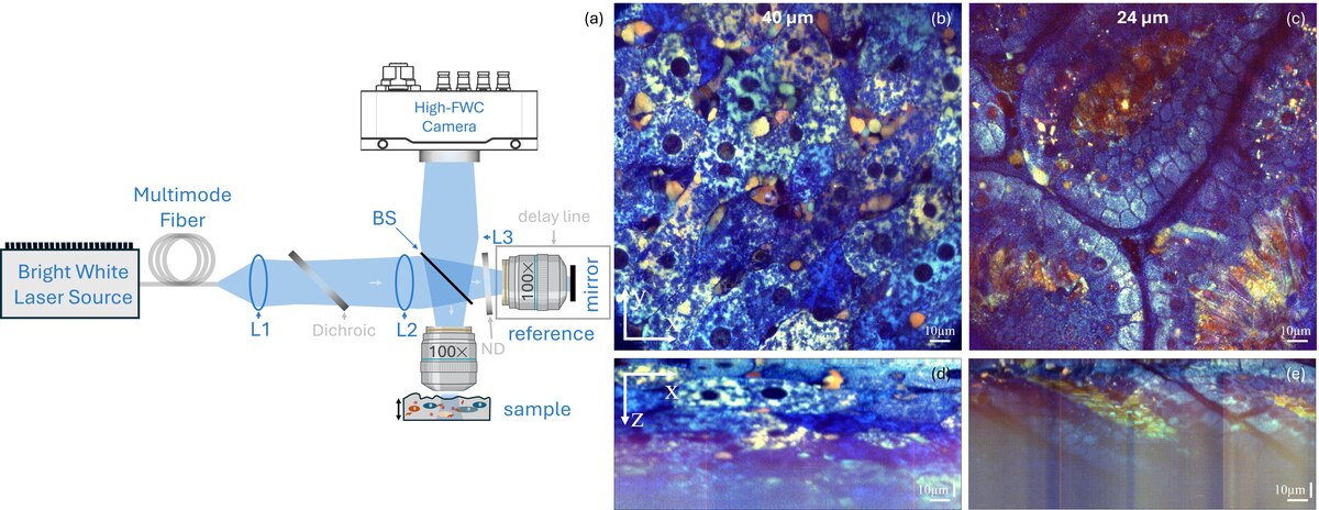
DYNAMIC FULL-FIELD OPTICAL COHERENCE MICROSCOPY FOR LIVE TISSUE IMAGING
Erikas Tarvydas1, 2, Austėja Trečiokaitė1, Egidijus Auksorius1
1 Center for Physical Sciences and Technology, Vilnius, Lithuania
2 Vilnius University, Faculty of Medicine, Vilnius, Lithuania
Acknowledgments: We thank Seemantini Nadkarni and Arvydas Laurinavičius for discussions, and Urtė Neniškyte, Viktorija Kralikienė and Igor Nagula for providing mouse tissues.
[1] J. Scholler et al., “Dynamic full-field optical coherence tomography: 3D live-imaging of retinal organoids,” Light Science & Applications, vol. 9, no. 1, p. 14.0, Aug. 2020, doi: 10.1038/s41377-020-00375-8
[2] K. Groux et al., “Dynamic full-field optical coherence tomography allows live imaging of retinal pigment epithelium stress model,” Communications Biology, vol. 5, no. 1, p. 575, Jun. 2022, doi: 10.1038/s42003-022-03479-6.
[3] E. Tarvydas, A. Treciokaite, and E. Auksorius, “High-Resolution Dynamic Full-Field Optical Coherence Microscopy: illuminating intracellular activity in deep tissue,” arXiv.org, Aug. 05, 2025. https://arxiv.org/abs/2508.03657