Pulse Electric Field (PEF) ablation is gaining significant clinical interest as a non-thermal alternative to traditional radiofrequency or cryotherapy-based catheter ablation for the treatment of cardiac arrhythmias. Despite PEF offering improved tissue selectivity and reduced collateral damage, reliable functional validation of lesion depth and electrical effectiveness remains essential for controlling clinical outcomes.
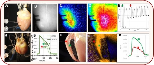
We introduce a multimodal cross-validated approach for functional assessment of PEF-induced ablation zones by combining high-speed optical mapping (OM) with microelectrode (ME) transverse and transmural action potential (AP) recordings (Fig.1). Experiments were conducted in a Langendorff-perfused rabbit heart with synchronized PEF delivery, cardiac pacing, and OM acquisition. Critically, lesion depth and electrical impairment were independently verified by recording APs in a cut-off ventricular wedge prepared from PEF-treated regions, enabling direct cross-validation of surface and transmural electrophysiological changes. Both single-pulse and complex PEF protocols were evaluated.
The results indicate that PEF significantly affects lesion size, depth, and functional severity. It slows AP propagation, causes conduction block, and alters AP morphology in peri-ablated tissue. Regions without electrical activity were identified at depths of up to 3 mm, depending on the stimulation parameters used. Overall, OM and ME techniques are useful for investigating and evaluating PEF ablation effectiveness.