Extracellular vesicles (EVs) contain biologically active molecules, such as microRNAs (miRNAs) and proteins, and have been emerging as a promising therapeutic strategy for neurodegenerative diseases [1]. One major advantage of EVs is their ability to cross the blood-brain barrier and efficiently deliver their cargo [2]. In this study, we investigated the molecular effects of EVs derived from human amniotic fluid stem/stromal cells (hAF-SCs) on SH-SY5Y neuronal cells that had reduced activity of the anti-neuroinflammatory microRNA miR146a-5p.
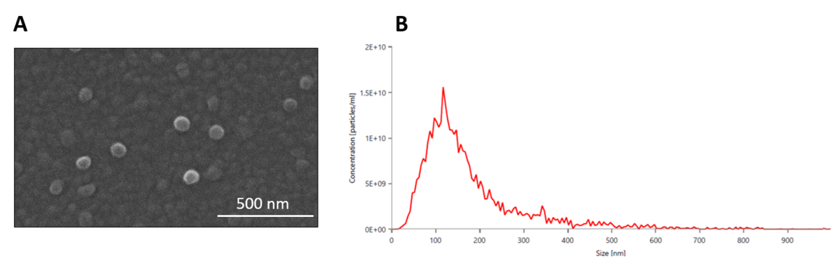
For this purpose, SH-SY5Y neuroblastoma cells were differentiated into a more mature neural phenotype using a specialized differentiation medium composed of BrainPhys™ neuronal medium supplemented with Penicillin/Streptomycin, serum- and antioxidant-free B-27™, 3.7 nM BDNF receptor agonist PG-003, and 10 µM retinoic acid. EVs were isolated from the hAF-SC conditioned media using size exclusion chromatography with qEVoriginal columns. The isolated EVs were characterized based on size using scanning electron microscopy (SEM) and nanoparticle tracking analysis (NTA) (Fig.1). Additionally, molecular markers were examined through western blotting (WB) and RT-qPCR. To inhibit the activity of miR146a-5p in the neuro-differentiated SH-SY5Y model, a mirVana inhibitor was utilized in combination with Lipofectamine RNAi/MAX transfection.
The results indicated that the most commonly detected particle size was 117.5 nm, and the EVs exhibited a round morphology. The extracted hAF-SC EVs were positive for typical EV markers, such as TSG101, CD63, and ALIX. Among the miRNAs tested, miR-16-5p, miR-146a-5p, and miR-210-3p showed the highest expression levels. Treatment of SH-SY5Y cells with 70,000 hAF-SC EVs per cell over 48 hours resulted in a significant increase in the expression of genes associated with neural processes, such as NTN3, MAGEL2, GABRD, SLURP1, P2RY4, and SIX1 as revealed by mRNA sequencing analysis.
In conclusion, these findings support the potential of hAF-SC EVs as a cell-free strategy for targeting neuronal molecular pathways relevant to neurodegenerative and neuroinflammatory conditions.