
The development of advanced transdermal therapeutic systems requires matrices that combine biocompatibility, mechanical integrity, and controlled release kinetics. This study focuses on the synthesis and characterization of multicomponent hydrogel films based on gelatin, polyvinyl alcohol (PVA), and hyaluronic acid (HA), crosslinked with glutaraldehyde. Gelatin provides essential biological signaling for cell adhesion and mimics the extracellular matrix, while PVA enhances the structural stability of the composite. The integration of HA, a high-molecular-weight anionic polysaccharide, aims to modulate the hydrophilicity and internal architecture of the polymer network to optimize drug diffusion. Rheological analysis of the Gelatin/PVA/HA compositions reveals non-Newtonian pseudoplastic behavior, where dynamic viscosity decreases as the shear rate increases. This characteristic is vital for the processing and application of biomedical gels. The introduction of HA significantly increases the initial viscosity in a concentration-dependent manner, indicating the formation of a more complex intermolecular network through hydrogen bonding between the HA functional groups and the base polymer chains. At high shear rates, the convergence of viscosity curves suggests the partial disruption of these physical interactions and a shift toward hydrodynamic dominance. Structural analysis through gel fraction determination shows that while glutaraldehyde effectively crosslinks the gelatin amino groups, HA remains primarily physically entrapped. 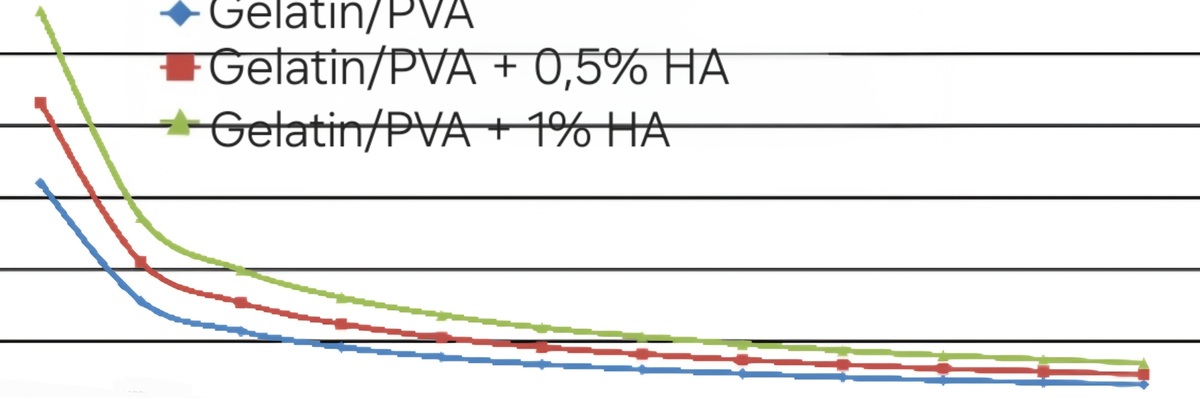
DEVELOPMENT AND RESEARCH OF THE PROPERTIES OF GELATIN-BASED HYDROGEL MATERIALS FOR TRANSDERMAL DRUG DELIVERY SYSTEMS
Daria Kuchynska1, Olena Ishchenko1, Ihor Okhrimenko1, Mikael Yeganyan1
1 Kyiv national university of technology and design, Department of industrial pharmacy, Kyiv, Ukraine
[1] K. J. Goudie, S. J. McCreath, J. A. Parkinson, C. M. Davidson, and J. J. Liggat, “Investigation of the influence of pH on the properties and morphology of gelatin hydrogels,” Journal of Polymer Science, vol. 61, no. 19, pp. 2316–2332, Jun. 2023, doi: 10.1002/pol.20230141.
[2] V. Dhote, S. Skaalure, U. Akalp, J. Roberts, S. J. Bryant, and F. J. Vernerey, “On the role of hydrogel structure and degradation in controlling the transport of cell-secreted matrix molecules for engineered cartilage,” Journal of the Mechanical Behavior of Biomedical Materials/Journal of Mechanical Behavior of Biomedical Materials, vol. 19, pp. 61–74, Nov. 2012, doi: 10.1016/j.jmbbm.2012.10.016.
[3] S. Bahmani, R. Khajavi, M. Ehsani, M. K. Rahimi, and M. R. Kalaee, “A development of a gelatin and sodium carboxymethyl cellulose hydrogel system for dual-release transdermal delivery of lidocaine hydrochloride,” International Journal of Biological Macromolecules, vol. 284, no. Pt 2, p. 138034, Nov. 2024, doi: 10.1016/j.ijbiomac.2024.138034.