
Surface-enhanced Raman spectroscopy (SERS) is an ultrasensitive vibrational spectroscopic technique that enables the detection of trace analyte concentrations while providing detailed molecular structure information. SERS employs nanoparticle-based substrates to enhance the Raman signal originating from molecules located at or near the surface. The enhancement arises from two main mechanisms: the electromagnetic mechanism, associated with localized surface plasmons, and the chemical mechanism, which involves direct interaction or binding of the molecule to the nanoparticle surface. Tuning excitation wavelength to the UV spectral range (e.g., 325 nm), known as UV-SERS, enables selective enhancement of Raman signals from biological molecules such as adenine or aromatic amino acids, even though plasmonic enhancement in the UV range is generally weaker. However, one of the key challenges in UV-SERS remains the production of chemically stable, nanostructured metallic substrates that provide sufficient enhancement in the UV range. For this purpose, nanoparticles can be produced by laser ablation in liquids, which ensures high chemical purity, and allows control over nanoparticle size and composition. In this work, 50 nm- and 100 nm-thick Cu, Au, Ag, and Fe films and their combinations were used to fabricate nanoparticles via laser ablation in acetone. These nanoparticles were then used for UV-SERS applications in biological molecule studies. The ability of these nanoparticles to enhance the Raman signal of adenine is analyzed, and their stability over time is evaluated in order to determine their suitability for long-term and repeatable measurements. 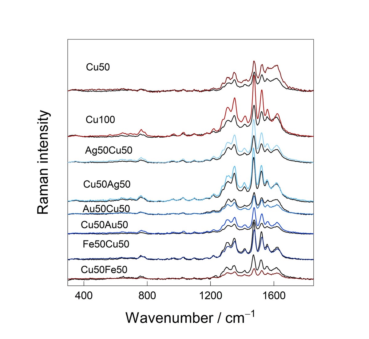
The study revealed that the strongest UV-SERS signal of adenine is obtained using copper nanoparticles from 100 nm thick Cu layer (Figure 1). After four months, the UV-SERS signal obtained from Cu₅₀Ag₅₀, Au₅₀Cu₅₀, and Fe₅₀Cu₅₀ nanoparticles remained unchanged or changed only slightly, demonstrating the highest stability among the investigated samples. Pure 50 and 100 nm iron, gold, and silver nanoparticles did not produce a detectable adenine spectrum using the UV-SERS method.