The second near-infrared spectral window (NIR-II, 1000-1700 nm) offers significant advantages in deep-tissue penetration, reduces photon scattering and minimizes tissue autofluorescence. These advantages require development of emissive nanomaterials with broad absorption, efficient near-infrared emission and high photostability. Polymer dots (Pdots) represent a promising class of probes for NIR-II bioimaging due to their high brightness, strong resistance to photobleaching and low cytotoxicity [1]. In this study, we investigate the optical and physicochemical properties of novel polymer dots and to evaluate their suitability as NIR-II fluorescent probes for bioimaging applications.
Polymer dots composed of the semiconducting polymer DBT-4O-TTQ and surface-modified with DSPE-mPEG (40%) were characterized following dilution in aqueous media and measurements of absorption and fluorescence spectra, photobleaching, zeta potential and particle size distribution.
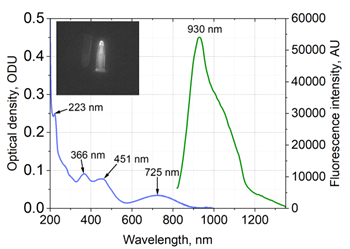
The Pdots exhibit broad absorption in the ultraviolet and visible regions, enabling efficient excitation at 808 nm. Under this excitation, a wide near-infrared emission band with a maximum at 930 nm was observed, extending toward longer wavelengths, relevant for NIR bioimaging (Fig. 1). Dynamic light scattering measurements reveal a hydrodynamic diameter of 85 nm with low polydispersity (PDI = 0.126), while zeta potential of -40 mV indicates good colloidal stability.
The acquired results indicate that Pdots combine broad spectral absorption, stable nanoscale morphology and near-infrared emission. The observed photophysical and optical properties highlights their potential as organic fluorescent probes for advanced bioimaging applications and motivates further NIR-II imaging studies in biological systems.