Diatoms are unicellular algae with a distinctive photosynthetic apparatus that enables them to adapt to extreme changes in light intensity [1]. This allows the cells to efficiently utilize available light for photosynthesis and determines photoprotection mechanisms. Cycclotella meneghiniana is a convienient model for diatom analysis. Cryogenic fluorescence spectroscopy is a valuable tool for studying electronic excitation dynamics in photosynthetic apparatus on the whole-cell level. A mixture of glycerol and growth media is commonly used in cryogenic measurements to reduce ice crystal formation, which causes light scattering. However, evidence supporting the viability of cells in glycerol is limited, as most conclusions are based solely on fluorescence spectra shape [2]. The aim of this study is to assess the effects of glycerol on C. meneghiniana cell viability and reveal the differences in spectral features of cells in different functional states measured at cryogenic temperatures.
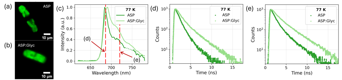
Cells were measured in either artificial seawater (ASP) or in medium for cryogenic measurements ASP:glycerol (2:3 ratio), and both steady-state fluorescence emission spectra and time-resolved fluorescence decays were measured at temperatures from 16K to room temperature (RT). Cell viability was assessed using propidium iodide, morphological changes in ASP and ASP:glycerol were assesed by fluorescence microscopy (Fig. 1a-b). Figure 1c-e contains representative data obtained at 77K. Diatoms exposed to ASP:glycerol exhibited rapid loss of viability, thylakoid membrane swelling (Fig. 1b), and longer fluorescence lifetimes, indicating disruption of the photosynthetic apparatus despite minimal changes in spectral shape. Medium exchange from ASP to ASP:glycerol led to significant changes in spectral features: the fluorescence spectrum peak position was red-shifted (Fig. 1c) and fluorescence lifetimes decreased, both for emission at 685 nm and 720 nm (Fig. 1d–e) in ASP:glycerol compared to ASP. In contrast, glycerol-free conditions preserved cellular integrity and produced a consistent, steadily changing fluorescence response from 16 K to 130 K (data not shown).
These results suggest that cryogenic studies using glycerol at the whole-cell level should be interpreted with caution, as cell viability needs to be confirmed.