HIGH-THROUGHPUT SPATIO-TEMPORAL OPTICAL COHERENCE TOMOGRAPHY FOR ENHANCED RETINAL IMAGING SENSITIVITY
Karolis Adomavičius1, Austėja Trečiokaitė1, Erikas Tarvydas1, Egidijus Auksorius1
1 Center for Physical Sciences and Technology, Optoelectronics department, Lithuania
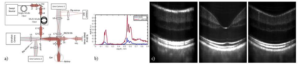
Optical Coherence Tomography (OCT) has rapidly become a crucial tool in ophthalmologic diagnostics. Nevertheless, traditional scanning systems are strongly limited by scanning speed, susceptibility to motion artefacts and optical aberrations. Spatio-Temporal Optical Coherence Tomography (S-TOCT) enables high-speed retinal imaging with computational aberration correction, but at the cost of reduced sensitivity. When using standard 50/50 beamsplitters, more than 75% of the available light is lost due to double losses: 50% of the light is lost during illumination, and another 50% is lost in signal collection. In addition, the reference reflectance must be kept low for biological tissue samples [1]—which further increases light losses. Simply increasing the source power is typically not viable due to source limitations or phototoxic effects on the sample. Here, we present a high-throughput (HT) STOC-T configuration that recovers a significant portion of the available light budget without increasing source power. The design replaces the standard 50/50 beamsplitter with an asymmetric 90/10 beamsplitter and incorporates a pupil-plane pick-off mirror [2], allowing illumination and signal detection through the same beamsplitter facet (Fig. 1a). This maximizes light delivery to the retina while efficiently collecting backscattered photons scattered over a wide angular range by retinal tissue.
Fig. 1. (a) Principal system used. Comparison of in vivo human retina imaging with standard (b, blue curve) and high throughput S-TOCT (b, red curve) acquired with line cameras. (c) Retinal cross-sections acquired with HT mode and 2D camera.
The HT-STOC-T system achieves up to a 3.5× improvement in sensitivity (≈5.5 dB) compared to a conventional STOC-T configuration, reaching sensitivities close to 100 dB with moderate volume averaging [3]. In vivo retinal imaging confirms these findings: axial S-TOCT images reveal layered structures that are visible only in the HT configuration (Fig. 1b, c). In vivo retinal imaging using a 2D ultrafast camera demonstrates the practical impact of this sensitivity gain. Figure 1c shows three retinal cross-sectional (XZ) images acquired in HT mode with ultrafast 2D camera and centered around the macular region [3]. Each image represents a distinct cross-section through the retina. All major retinal layers—including the nerve fiber layer, inner and outer plexiform layers, the retinal pigment epithelium, and the choroid—can be clearly identified across the images, indicating improved depth-resolved contrast and signal quality throughout the retinal volume.
By substantially improving sensitivity while preserving ultrafast volumetric acquisition and computational aberration correction, the high-throughput STOC-T configuration enhances the capability of STOC-T for in vivo retinal imaging and low-signal biological applications.
[1] A. Dubois, L. Vabre, A.-C. Boccara, and E. Beaurepaire, “High-resolution full-field optical coherence tomography with a Linnik microscope,” Applied Optics, vol. 41, no. 4, p. 805, Feb. 2002, doi: 10.1364/ao.41.000805.
[2] E. Auksorius and A. C. Boccara, “High-throughput dark-field full-field optical coherence tomography,” Optics Letters, vol. 45, no. 2, p. 455, Dec. 2019, doi: 10.1364/ol.381888.
[3] K. Adomavičius, A. Trečiokaitė, E. Tarvydas, and E. Auksorius, “High-throughput spatio-temporal optical coherence tomography (STOC-T),” Optics Continuum, vol. 4, no. 9, p. 2099, Aug. 2025, doi: 10.1364/optcon.564109.