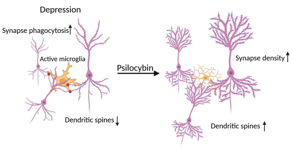
According to the World Health Organization, Major Depressive Disorder (MDD) affects 5.7% of adults, of which 30% have treatment-resistant depression; therefore psychedelics are being investigated as a potential new therapy. MDD presents itself with a decrease in brain structure volume and plasticity, especially prefrontal cortex, hippocampus and amygdala [1]. Microglial cells are brain immune cells, that also prune inactive synapses. It is also known that in MDD, microglial phagocytosis of synapses increases, which leads to reduced synaptic plasticity. Psilocybin is now being researched as a possible treatment for MDD since it increases neuroplasticity and alleviates depression symptoms [2]; however, the mechanism is still not known. It is known that psilocybin lowers microglial phagocytic activity and production of ROS and NO [2], [3]. We hypothesize that psilocybin induces the number of synapses by modulating microglial activity (Fig. 1). This study aims to compare the number of synapses in stressed and psilocybin-treated animals and to identify microglial involvement in this process.
Experiment included six mice groups: control, 1 mg/kg psilocybin, 2 mg/kg psilocybin, held while others experienced CDM and saline, stressed and saline, stressed and 1mg/kg psilocybin. The brains were cryosectioned and stained using immunofluorescence. To identify synapses, we used presynapse (anti-VGAT) and postsynapse (anti-GEPHYRIN) antibodies and microglial (anti-IBA1) in the dorsal hippocampus. Brains were imaged using the SP8 confocal microscope. The differences in synaptic density between groups are presented in 3D reconstruction images obtained using IMARIS software.
Understanding the mechanism of psilocybin-induced neuroplasticity could help understand microglia cell physiology and extend MDD treatment options.