Increasing use of nanoparticles (NPs) in industry and biomedicine raises new environmental safety challenges. Assessing NPs cytotoxicity is critical for developing safe technologies, particularly for quantum dots (QDs) - photoluminescent semiconductor NPs, that can interact with primary producers like algae. Although cadmium-free QDs are emerging as safer alternatives in biomedicine, their impact on photosynthetic organisms remains poorly understood. Additionaly, the role of an organism’s physiological state in mediating QD-induced stress is often overlooked. This study examines how differently photoadapted algal cultures respond to Cu-based QDs, revealing links between environmental adaptation and NPs effects.
In this study, two Desmodesmus communis algae cultures (from Nature Research Center) were analyzed. Both were transitioned from high-light (100 μmol photons/m\(^{2}\)s) to lower-light conditions (34 μmol photons/m\(^{2}\)s, 12/12 h day/night photoperiod, MWC growth media) for different durations: one week (Culture No.1) and six months (Culture No.2), establishing distinct photoadaptation states. Samples were exposed to 4 μM CuInZnS/ZnS (Nanooptical materials, USA) QDs for 24 hours. The physiological impact and pigment dynamics were assessed using non-invasive in vivo techniques, including UV-VIS and PAM fluorometry, alongside fluorescence and Coherent anti-Stokes Raman (CARS) microscopy.
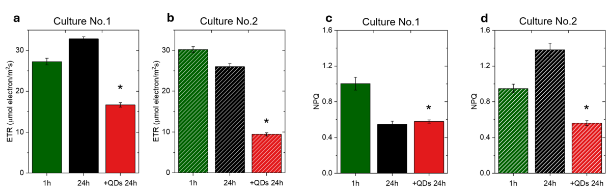
Our results demonstrate that although exposure to QDs generally causes disruption of energy distribution in PSII, as evidenced by decreased electron transport rate (ETR) and impaired non-photochemical fluorescence quenching coefficient (NPQ) in both cultures (Fig. 1), the nature of the resulting damage largely depends on the physiological state of the algae cells. In Culture No.1 (retaining high-light traits), this energetic imbalance quickly progressed to irreversible cellular damage, as evidenced by pigment degradation and pronounced alterations in autofluorescence as well as CARS data. In contrast, Culture No.2 (fully low-light adapted), primarily exhibited functional failure (registered sharp decrease in ETR) despite a more stable morphology. CARS microscopy further indicates that impaired photoprotection in the latter culture may be associated with functional uncoupling of carotenoids from their quenching complexes, leading to their locally enhanced vibrational visibility.
In conclusion, this study demonstrates that photoadaptation dictates whether QDs-induced stress leads to cellular degradation or functional impairment, providing a new mechanistic understanding of how NPs interact with the photosynthetic apparatus of algae.