
Surface-enhanced Raman spectroscopy (SERS) provides chemically specific molecular fingerprint information with sensitivity reaching the single-molecule level. Most SERS studies employ visible or near-infrared excitation; however, in these spectral regions fluorescence background and overlapping vibrational bands can reduce spectral contrast and limit discrimination between closely related biomolecules. In biomedical applications, these limitations are further compounded by substrate heterogeneity and the presence of complex biological matrices. Ultraviolet SERS (UV-SERS) addresses these constraints by exploiting the strong electronic absorption of aromatic biomolecules in the ultraviolet region, which selectively amplifies Raman scattering and improves molecular selectivity (Fig. 1). Many biologically and analytically relevant low-molecular-mass aromatics (nucleobases, aromatic amino acids, flavins, and numerous dyes) have strong electronic transitions in the UV. Thus, excitation at 229–325 nm can place these analytes under resonance or pre-resonance Raman conditions, producing strong, mode-selective amplification of vibrational bands that is not obtained for non-UV-active species. This is explicitly evidenced in your multiwavelength copper study, where π-conjugated analytes (adenine, guanine, riboflavin, 4-mercaptobenzoic acid) show pronounced UV and deep-UV enhancement, whereas an analyte lacking an extended π system (1-dodecanthiol) shows no vibrational signature bands, supporting the selectivity argument [1]. 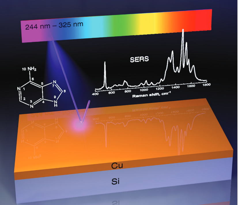
UV-SERS ON Cu, Co, AND Pd NANOSTRUCTURES FOR SELECTIVE DETECTION OF SMALL AROMATIC BIOMOLECULES
Martynas Talaikis1, Evaldas Stankevičius1, Rodrigas Liudvinavičius1, Lina Mikoliūnaitė1, Aikaterini-Maria Gkouzi1, Shivani Yadav2, Vladimir Sivakov2, Gediminas Niaura1
1 Center for Physical Sciences and Technology, Vilnius, Lithuania
2 Leibniz Institute of Photonic Technology, Jena, Germany
Acknowledgments: This work was supported by the Research Council of Lithuania (LMTLT) under an agreement number S-MIP-23-30 “Magneto-plasmonic nanoparticles for UV-SERRS detection of low-molecular-weight biomarkers (MAGUV-SERRS)”.
[1] S. Yadav, M. Talaikis, Y. V. Ryabchikov, M. Ziegler, G. Niaura, T. Deckert-Gaudig, V. Sivakov. “Copper-Based Multiwavelength Ultraviolet Surface -Enhanced Resonance Raman Spectroscopy.” Advanced Optical Materials 2025, 13, 2500078. https://doi.org/10.1002/adom.202500078
[2] M. Talaikis, R. Liudvinavičius, E. Stankevičius, A. Selskienė, A. M. Gkouzi, T. Murauskas, V. Sivakov, G. Niaura. “Femtosecond Laser-Induced Nanostructures in Copper Film for UV-SERS.” ACS Applied Materials & Interfaces, 2026, acsami.5c21582. https://doi.org/10.1021/acsami.5c21582.