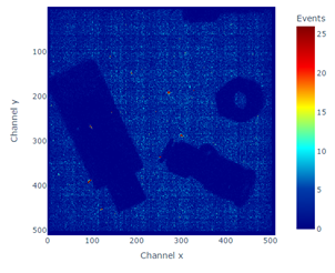
A fully integrated Gas Electron Multiplier (GEM) detector system was used for high‑resolution X‑ray imaging. The detector employs a triple‑GEM foil structure with a 100 cm² active area enclosed in a sealed gas chamber. Each copper GEM foil contains a dense matrix of identical microscopic holes that enable electron avalanche multiplication under applied high voltage. Using a triple‑GEM configuration provides high gain, low ion backflow, and excellent spatial resolution.
The detector features a Kapton entrance window and a drift cathode that generate a uniform electric field across the drift region, guiding primary ionization electrons toward the GEM stack. After amplification through the three foils, the electrons are collected on a readout plate, yielding accurate spatial information about the incident radiation.
Signal processing is performed by a custom 256‑channel readout board developed by Techtra and connected to the detector via four Panasonic connectors. The board incorporates four Texas Instruments DDC264 ADCs, each providing 64 current‑input channels with 20‑bit resolution. Adjustable full‑scale charge ranges from 12.5 pC to 150 pC and integration times between 160 µs and 1 s allow precise current measurement across a wide dynamic range.
The analog front end includes precision reference sources, low‑pass filtering, and a biasing circuit capable of supplying a constant current to support negative‑polarity input signals. A Xilinx FPGA manages timing, data acquisition, and communication. Digitized data are serialized and transferred to a PC over a 100 Mbit/s Ethernet interface, where TCP is used for system control and UDP for high‑rate data streaming. The system operates at a 6.25 kHz sampling rate and is supported by software enabling real‑time monitoring, visualization, and data storage.
The detector operates with an Ar/CO₂ (70/30) gas mixture and is powered by a Caen DT5470N high‑voltage supply capable of delivering up to −5 kV at 200 µA. High voltage is routed to the GEM foils through a dedicated connector and cable. The assembly is housed in a gas‑tight enclosure equipped with interfaces for gas inlet and outlet, HV distribution, and signal readout, ensuring stable performance within controlled gas condition.
To evaluate the detector’s imaging performance, a COOL‑X miniature X‑ray source was used. This solid‑state device employs a pyroelectric crystal to generate electrons, which then produce X‑rays when accelerated into a copper target. The source delivers peak intensities of approximately 10⁸ photons per second, with endpoint energies reaching up to 35 keV, providing a compact and reliable excitation source for characterization of the GEM‑based imaging system.