
Enamel microcracks can contribute to tooth decay, reduced mechanical strength, and may act as precursors to larger structural fractures\(^{1}\). Accurate characterization of these microcracks using micro-computed tomography (µCT) and scanning electron microscopy (SEM) depends on careful sample preparation, including controlled dehydration. Improper dehydration can introduce artifacts such as contamination and dehydration-induced crack formation or enlargement, thereby compromising the reliability of microstructural analysis\(^{2}\).
Although extracted teeth are widely used in µCT\(^{1,2}\) and SEM studies\(^{3,4}\), dehydration protocols are often insufficiently reported or systematically assessed. A systematic literature review identified 70 relevant articles published within the last decade; 31 studies passed initial screening, and only 4 met the final inclusion criteria, corresponding to ~6% of the initially identified literature.
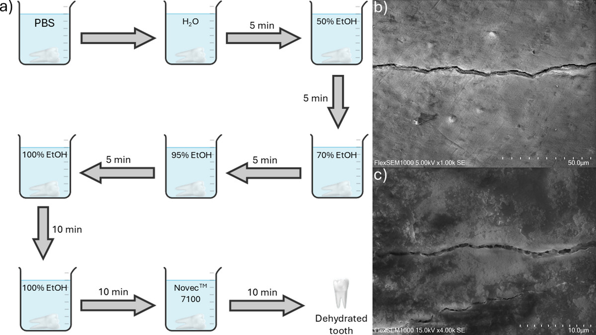
Teeth are commonly stored in phosphate-buffered saline (PBS) to maintain chemical and physical stability prior to analysis. Water present within microscopic pores, interprismatic spaces, and microcracks can generate capillary stresses during rapid evaporation that may exceed the local mechanical strength of enamel, promoting crack propagation, particularly during vacuum-based analyses such as SEM. Based on these considerations, we present an experimental tooth dehydration protocol (Fig. 1 a) involving stepwise substitution of water with increasing ethanol concentrations, followed by immersion in a low–surface-tension liquid (NovecTM 7100) prior to air drying.
In conclusion, tooth storage and dehydration conditions are frequently underreported and insufficiently controlled in µCT and SEM studies, limiting data reliability and cross-study comparability. The proposed dehydration protocol provides a standardized and explicitly documented framework that minimizes dehydration-induced artifacts and supports preservation of native enamel microstructure for reliable microcrack analysis (Fig. 1 b, c).