
Optical coherence tomography (OCT) is an interferometric imaging method – widely used for retinal imaging – that can image deep in tissue. Full-field OCT (FF-OCT), compared to classical OCT systems, uses a CMOS camera instead of a single point detector, which eliminates the need to scan sample in both x and y directions, and achieves higher lateral resolution [1]. Moreover, if the fastest CMOS camera is employed and a Fourier-domain data acquisition method is used (by utilizing a tunable narrowband light source) FF-OCT becomes a powerful tool, suitable for high-speed eye imaging [2]. However, crosstalk formation in images has been limiting the technique's performance preventing from seeing choroid in the human retina.
Choroid is a crucial determinant in the pathogenesis of many ocular diseases of the posterior pole as its principle role is to supply oxygen to the outer retina [3]. Crosstalk noise appears due to the use of the wide-field detection and spatially coherent laser, which can compromise image quality effectively reducing its spatial resolution, especially deep in tissue. A spatially incoherent light source can be used to remove crosstalk, as it makes laterally scattered photons that are detected in other pixels average to uniformity. Therefore, we have previously solved the crosstalk problem by reducing spatial coherence of the laser by the help of an ultrafast deformable membrane [4]. We demonstrate that a multimode fiber with carefully chosen parameters [5] enables crosstalk-free imaging over long axial range and without significant artifacts. Specifically, we study here how depth of field (DOF) is extended because of the use of the multimode fiber. Finally, we show that it can be used to image the human retina and choroid in vivo with exceptional contrast, as shown in Fig. 1.
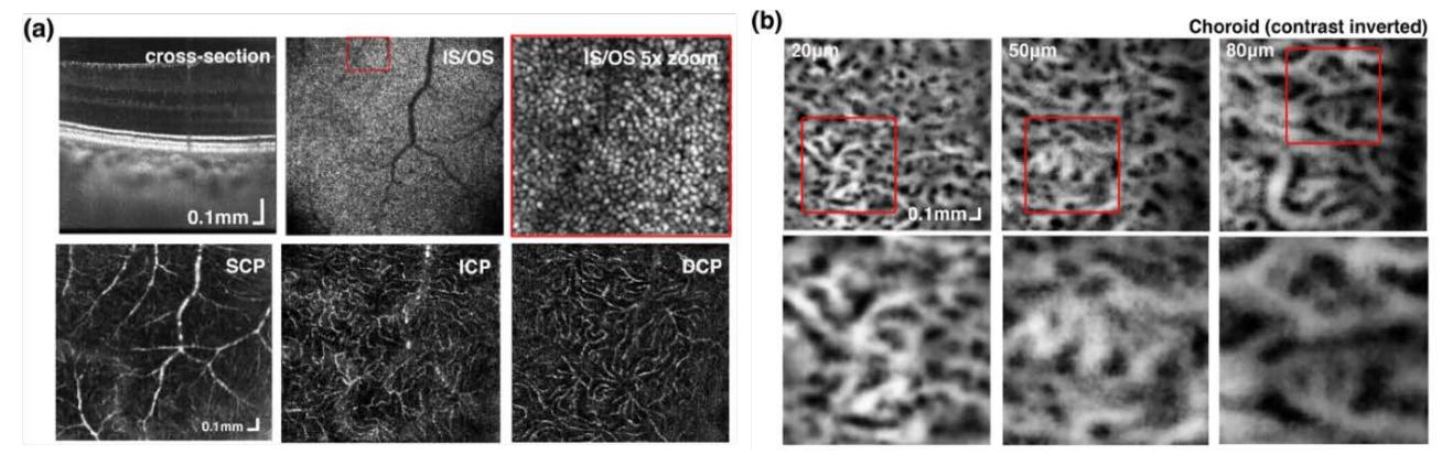
In conclusion, FD-FF-OCT as a tool for high-speed eye imaging has limitations due to crosstalk and coherence noise. However, we have demonstrated that carefully selected multimode fiber parameters and Fourier space filtering can produce crosstalk-free high-resolution images.