
Urolithiasis is a disease of stone formation in urinary tract. Most commonly occurring stones are calcium oxalate stones, other types of stones such as uric acid, struvite, calcium phosphate and cystine are found in urinary tract but less frequently than the calcium oxalate ones.
It is widely accepted that infrared (IR) spectroscopy is well suited for the identification of chemical composition of urinary stones, and some libraries of IR absorption and IR ATR (attenuated total reflection) spectra are available. The urinary sediments also can be analyzed by means of infrared spectroscopy, but this research field is still underdeveloped due to the sampling problems related to extremally small size of the sediments (deposits). The main task of this study is to extend capabilities of the IR spectroscopy to early diagnostics using urine samples.
Infrared spectroscopic study of deposits of patient urine could be a simple and non-invasive way to detect urinary stone formation and determine its composition. This could help in early detection of the stone formation of certain type for patient with suspected stone disease. Knowledge about chemical composition of the stone allows to prescribe specific medicines or diet to prevent further formation of stones. We have performed pilot studies of the set of samples removed from various patients. The set consists of three parts – urine before medical surgery (A), during the surgery (B) and removed urinary stone (C). Urine samples were first centrifuged to get urinary deposits, then deposits were measured using an ATR FTIR spectrometer. IR absorption spectra of the removed urinary stones were also measured.
From 26 patients, in 11 cases sample A only had organic compounds, no characteristic chemical components of the stones were detected. Among the remaining 15 cases, 13 IR spectra of A samples well correlated (86.7%) with the spectra of the removed stones. Our spectroscopic pilot study leads to conclusion that IR absorption spectra of urine deposits (small crystals) allows us to diagnose urolithiasis and to determine chemical composition of the stones with accuracy up to 86.7%.
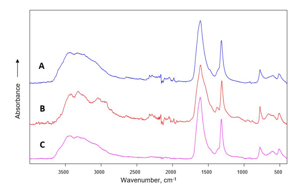
From 26 patients, 3 samples B had no characteristic chemical components of the stones, only organic compounds were deposited. Among the remaining 23 cases, 22 B samples the IR spectra correlated with removed stones spectra with accuracy of 95.7%. Such correlation could be helpful since endoscopic surgeries without complete removal of the stones became widely used. In such circumstances measuring the IR spectra of deposits of patient urine could be useful to determine composition of the stones and help patients prevent further formation of the stones by recommending specific diet and medication.