Protein aggregation is a biological phenomenon in which intrinsically disordered or misfolded proteins aggregate and form so-called amyloid fibrils characterized by fibrillar morphology. In living organisms, misfolded protein aggregates are hallmarks of various amyloidosis [1]. Several common neurodegenerative diseases such as Alzheimer's, Parkinson's or Huntington's disease are induced by the formation of pathogenic amyloid fibrils in living tissues. An important agent in the formation of amyloid fibrils is the destabilization of the native conformation of the protein. This process can be caused in vitro by the change of certain parameters, including pH, temperature, centrifugation, salinity or seeding. However, many proteins are organized naturally in the form of fibrils, having different structures and regulatory functions. The morphological and structural plurality of the protein fibrils and their toxicity are ambiguous, so better understanding of the supramolecular organization of amyloid fibrils may help in the treatment of many diseases.
Chiroptical spectroscopies, such as VCD (Vibrational Circular Dichroism) and ECD (Electronic Circular Dichroism), which are sensitive to chiral biological systems, were used in this study to show variations in the structure of obtained amyloid fibrils at different levels of their hierarchy. Due to a regular long-range chiral organization of amyloid fibrils, it was possible to observe the enhanced VCD signal [2]. By the appropriate selection of the obtaining parameters, we were able to obtain fibrils with controlled reversal of supramolecular helical chirality.
Moreover, ECD information about the secondary structure changes during protein aggregation process. Additionally, to establish the morphology of amyloid fibrils, TEM (Transmission Electron Microscopy) was applied.
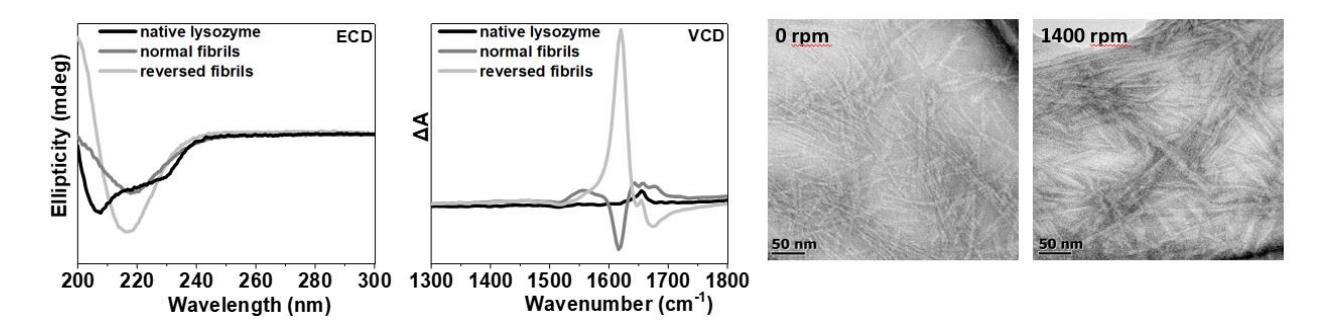
The results of VCD and ECD measurements of hen egg white lysozyme are presented in Figure 1. ECD spectra confirm aggregation process (fibrilization), as a transition of the $\alpha$-helix into $\beta$-sheet secondary structure is observed. VCD spectra show negative bands originated from the normal fibrils and intense positive signal typical for reversed fibrils, obtained due to intensive agitation (1400 rpm). TEM images present different morphology of the normal and reversed fibrils. It can be noticed that there are visible differences between normal and reversed fibrils not only in their structure, but also in their morphology. To summarize, the research enabled us to obtain fibrils of tailored structures due to proper choice of fibrilization parameters and monitoring of the structures via chiroptical spectroscopies.