
The emergence of multidrug resistance amongst nosocomial infection agents is one of the most pressing worldwide health issues. One of the ways to combat biofilm-associated bacterial infections is antimicrobial photodynamic therapy (aPDT). aPDT is based on the interaction of a photosensitizer, the appropriate wavelength of light and oxygen. After light excitation, the triplet-state photosensitizer interacts with molecular oxygen, electron donors or acceptors and can produce reactive oxygen species, thereby triggering photo-oxidative reactions that initiate various cellular damages and destroy the bacterial cells. [1]
Stenotrophomonas maltophilia is an emerging global multidrug-resistant opportunistic pathogen, which resistance to antibiotics is associated with a biofilm formation [2]. There is no published data about the aPDT caused inactivation of S. maltophilia pathogen. The main goal of the present study was to investigate the inactivation of S. maltophilia by riboflavin-based (RF-aPDT) and chlorophyllin-based aPDT (Chl-aPDT). The LEDs were chosen as optimal for the selected PSs (402 nm and 440 nm for Chl and RF, respectively) Two multidrug-resistant biofilms forming clinical S. maltophilia isolates (SM3 and SM21) were used for aPDT [3]. Bacterial cultures (~1×10⁷ CFU/mL) were suspended with 0.011 mM RF or 0.015 mM Chl in the dark. For the photoinactivation, 200 μl of the samples were exposed to light for different periods. The antibacterial effects of aPDT were evaluated by the microdilution method. The surviving cell populations were counted and the results were expressed as log10 of CFU/mL.
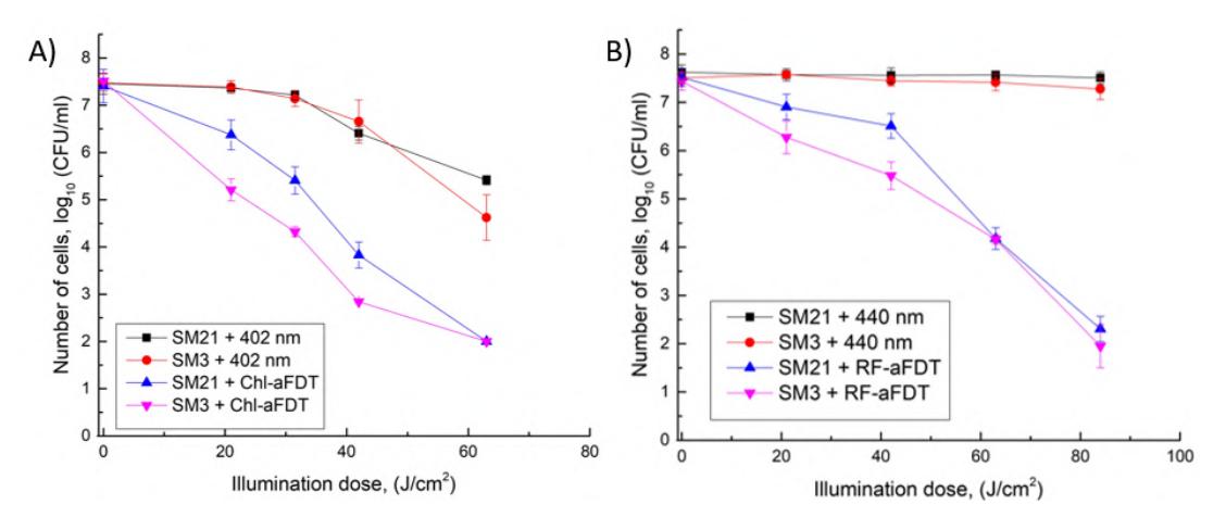
The experiment results showed that S. maltophilia SM3 isolate was more sensitive to RF-aPDT when the illumination doses were 21 J/cm2 and 42 J/cm2 compared to SM21. The increase of illumination dose to 84 J/cm2 led to similar sensitivity, where the viability of SM3 and SM21 after RF-aPDT were diminished by 5.5 log10 and 5.1 log10, respectively (Fig. 1A). The SM3 isolate was more susceptible to Chl-aPDT (reduction by 4.6 log10) than SM21 (reduction by 3.4 log10) when the illumination dose was 42 J/cm2 (Fig. 1B). Nevertheless, 402 nm LED light without PS (63 J/cm2) reduced survival of SM3 and SM21, CFU number decreased by 100 and 800 times respectively, whereas 450 nm light had no impact on both isolates. When illumination dose is 63 J/cm2, Chl-aPDT more sufficiently damages S. maltophilia cells than RF-aPDT. In summary, RF-aPDT and Chl-aPDT can be used as a potential means for S. maltophilia inactivation.