
Bacteriorhodopsin is a protein responsible for ion transfer across cell membrane.[1,2] To move ions light energy is required.[1,2] The active center of the bacteriorhodopsin is retinal.[1] When retinal absorbs light energy, its configuration changes from all-trans to 13-cis, 15-anti.[1] These changes initiates proton transfer process.[1] After some time, retinal configuration returns to its initial state, and proton transfer process may be repeated.[1] This process is called bacteriorhodopsin photocycle.[1,2]
It is known that retinal's 13-cis, 15-anti configuration duration is in the order of ps, μs, but ab-initio molecular dynamics (MD) calculations show that retinal's 13-cis, 15-anti configuration duration is in the order of fs.[2,3] Our main objective is to evaluate stability of retinal's 13-cis, 15-anti configuration.
In this work, we used Gaussian16 package. Calculation parameters: functional B3LYP, and basis set cc-pVDZ. For ab-initio molecular dynamics we used ADMP method. A lot of studies use these methods for bacteriorhodopsin modeling, because they describe bacteriorhodopsin protein quite well.[4]
We took 2 bacteriorhodopsin structures from pdb bank: 1C3W (ground state bacteriorhodopsin), and 2NTW (L state bacteriorhodopsin). From both structures, we cut out retinal, added hydrogen to retinal, and run structure optimization. Then we examined how environment affects 13-cis, 15-anti configuration stability by adding residues: Trp86, Trp182, Tyr185, and Thr90 to L state system, optimizing active center, and running MD calculations. After that we investigated which part of retinal's Schiff base, and Lys216 rotates when configuration changes occur, by rotating specific parts of Schiff base, and Lys216, and observing energy changes. Then we monitored how additional structures affects 13-cis, 15-anti configuration stability by adding more residues: Phe219, Leu93, Val20, and Met20 to our L state system, optimizing active center, and running MD calculations. Finally, we investigated 13-cis, 15-anti configuration stability by adding additional structures: Asp85, HOH401, HOH406 to L state system, freezing all atoms, except few, then running structure optimization, and observing configuration changes.
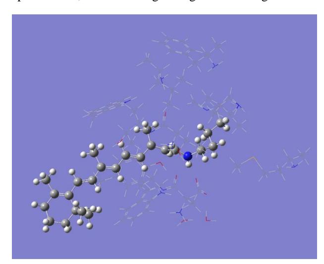
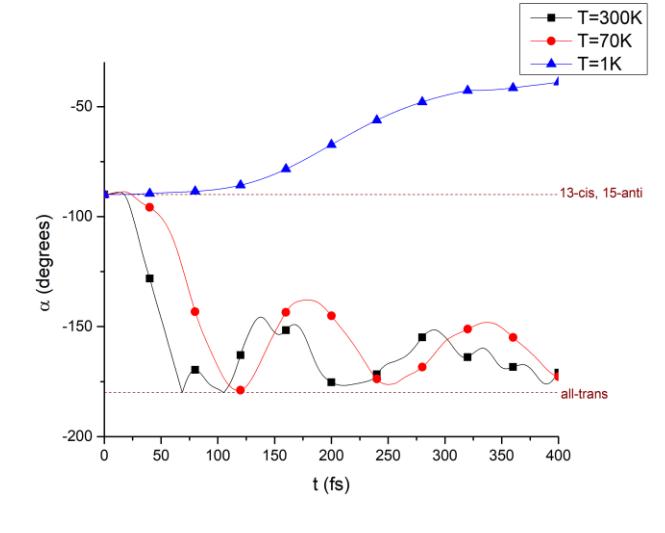
MD calculations show, that retinal's 13-cis, 15-anti configuration duration in vacuum is $t = 30$ fs at $T = 1$ K, and with residues from protein: Trp86, Trp182, Tyr185, and Thr90 is $t = 130$ fs at $T = 1$ K. When we added residues: Phe219, Leu93, Val20, and Met20 duration of 13-cis, 15-anti configuration became $t = 60$ fs at $T = 1$ K. This work final conclusion is that stability of configuration depends on residues from protein, and right residues must be recognized in order to increase stability of configuration.
Computations were performed on resources at the supercomputer "VU HPC" Saulėtekis of Vilnius University in Faculty of Physics.