
Obesity and obesity-related comorbidities including hypertension, diabetes or cardiovascular diseases pose a significant health problem in developed and developing countries. In obesity, the adipose tissue rapidly expands and its functions become disrupted, leading to inflammation [1]. Carotenoids, such as astaxanthin (AXT), are among natural compounds that show a protective effect against inflammation [2]. AXT is a widespread carotenoid, present especially in many aquatic organisms, such as salmon or shrimps [3]. AXT as a superpotent antioxidant and an anti-inflammatory agent may be an important factor in prevention and treatment of chronic diseases and related conditions [2].
AXT molecule consists of 40 carbon atoms which construct a polyene chain and two terminal ionone rings. The presence of chromophore–conjugated double bonds and two chiral centers gives AXT its distinctive spectroscopic properties and makes it an interesting subject for chiroptical studies [2]. Within the organism AXT is transported in a bound state, forming complexes with plasma proteins [4]. Due to its lipophilic properties, AXT binds to lipid membranes and is stored mainly in lipid droplets of adipocytes [5].
In this work, we use AXT chiral properties to observe changes in the carotenoid structure occurring due to its binding to bovine serum albumin (BSA). ECD spectra of AXT in tetrahydrofuran (THF) and conjugated to BSA in phosphate-buffered saline (PBS) (Fig. 1) show significant changes in AXT structure in the presence of protein.
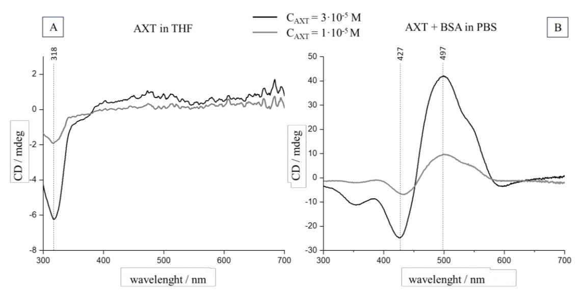
The analysis of obtained spectra indicates that AXT binds to BSA forming aggregates, which is visible as an appearance of the exciton couplet in the ECD spectra (Fig.1B). We presume that the binding of astaxanthin to serum protein may facilitate carotenoid transport to adipocytes, which is going to be examined using Raman microscopy.