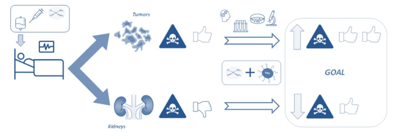
Cisplatin is one of the most widely used antitumor drugs in the world, but its clinical use is limited by the side effects that it could produce in patients, such as nephrotoxicity [1]. This has motivated, for decades, an extensive search for renoprotective strategies that allow improving the effectiveness of the drug, which is still irreplaceable in many cases [2].
One possibility is the use of selenium nanoparticles (SeNPs), the object of research for various current biomedical applications [3]. Therefore, in this work we tested the feasibility of SeNPs as a potential nephroprotective agent in treatments with cisplatin, due to the intrinsic antioxidant properties of selenium, which could reduce oxidative damage in kidney cells.
To consider a possible nephroprotective capacity of the SeNPs, they should not only protect renal cells against the cytotoxicity of cisplatin, but at the same time it would be essential that they do not interfere with the anticancer action. Hence, as a first approach to this purpose, in the present work HeLa cells (from cervical cancer) were cultured at different incubation times with cisplatin and/or with chitosan-stabilized SeNPs (Ch-SeNPs).
Cell viability was determined by the MTT assay ((3-(4,5-dimethylthiazol-2-yl)-2,5-diphenyltetrazolium bromide) tetrazolium reduction assay) for all cultures. The morphology and size of the Ch-SeNPs, as well as their cellular internalization, were studied using TEM-XEDS (Transmission Electron Microscopy coupled to Energy-Dispersive X-ray Spectroscopy). The intracellular content of Se and Pt was quantified by ICP-MS (Inductively Coupled Plasma Mass Spectrometry) analysis, after acid digestion of the cell pellets, and also the amount of Pt bound to DNA, which is the final target of the drug. Furthermore, Se and Pt speciation studies were carried out at the cellular cytosolic level, using liquid chromatography (LC) coupled to ICP-MS or UV/VIS.
The results showed that cells could assimilate these SeNPs, which were found in different subcellular locations, but their coadministration with cisplatin had no influence on the cellular drug uptake or the quantity of DNA-bound platinum, two factors traditionally related to Pt-drug cytotoxicity. However, in terms of cell viability, HeLa suffered more damage when cisplatin was mixed with SeNPs than in the case it was administered alone, suggesting a possible synergistic effect, which could be interesting in order to try to decrease cisplatin clinical dose and also the risk of nephrotoxicity.
Although this work is preliminary and it should be extended to kidney cells, the above results are promising enough for further study of the applications of SeNPs against renal toxicity in antitumor treatments with cisplatin.