
Acinetobacter baumannii and Stenotrophomonas maltophilia are multidrug resistant opportunistic pathogens, with most virulent strains found in hospitals, and are among the main causes of nosocomial outbreaks.[1][2] New preventative measures against these bacteria are needed because of their ability to quickly gain antibiotic resistance through horizontal gene transfer or spontaneous mutations. One of the perspective methods is antimicrobial photodynamic therapy (aPDT) – a biophotonic technology, where a photosensitizer is excited at an appropriate wavelength and inactivates microorganisms by creating reactive oxygen species in the presence of molecular oxygen.[3]
5-aminolevulinic acid (ALA) is a porphyrin precursor in the heme biosynthesis pathway. An excess of exogenously added ALA in bacterial cells leads to accumulation of endogenous porphyrins, such as coproporphyrin, uroporphyrin and protoporphyrin IX, which can act as photosensitizers for aPDT when excited within the Soret band spectral range.[4]The aim of this study is to find out whether A. baumannii and S. maltophilia are capable of producing endogenous porphyrins when incubated with 1 mM ALA and to assess their arrangement in intracellular and extracellular environments.
We evaluated the synthesis of porphyrins in bacteria (~1×10⁷ colony forming units per milliliter) at different incubation periods (0 – 24 h) with 1 mM ALA by measuring fluorescence intensity at excitation wavelengths within the Soret band (390 nm and 405 nm). Extracellular porphyrin fluorescence spectra were recorded after filtrating the solution through 0.22 μm filter. Fluorescence excitation and emission spectra were recorded by means of a LS55 fluorescence spectrophotometer (PerkinElmer, USA). Excitation and emission slits were set to 5 and 10 nm, respectively.
The results indicate that the amount of porphyrins increases over time in both bacteria incubated with ALA as seen from the fluorescecne intensity being monitored at excitation wavelength 390 nm (not shown) and 405 nm (Fig. 1). Compared to A. baumannii, S. maltophilia generates a greater amount of porphyrins at λex.= 390 nm (not shown). At excitation wavelength of 405 nm, fluorescence bands of ALA-induced porphyrins synthesized by A. baumannii shift towards 618 nm, which is characteristic of uroporhyrin (Fig. 1 A). In contrast, the emission spectra of S. maltophilia shift towards 612 nm, which corresponds to coproporphyrin (Fig. 1 B).
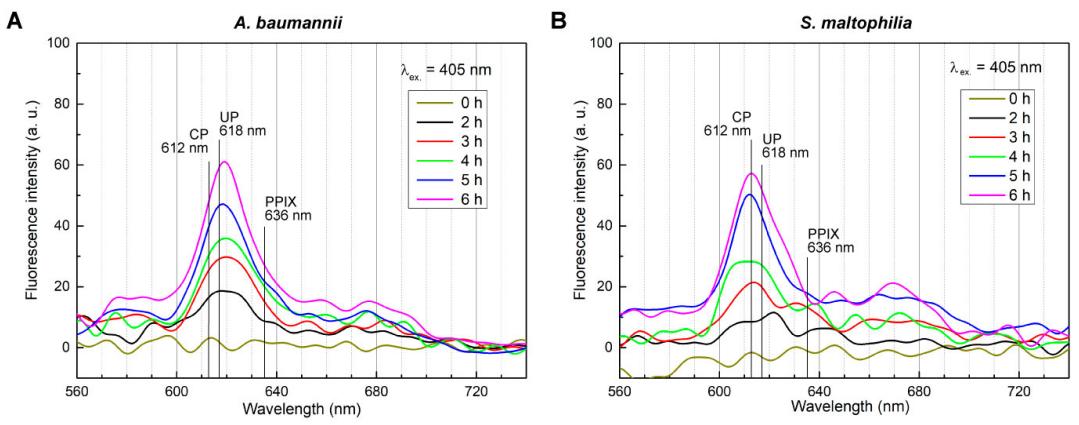
In conclusion, both A. baumannii and S. maltophilia are capable of synthesizing porphyrins when incubated with externally added ALA. Part of them bacteria excrete to extracellular environment, while the rest stays in intracellular environment (data not shown) which makes them suitable for aPDT.