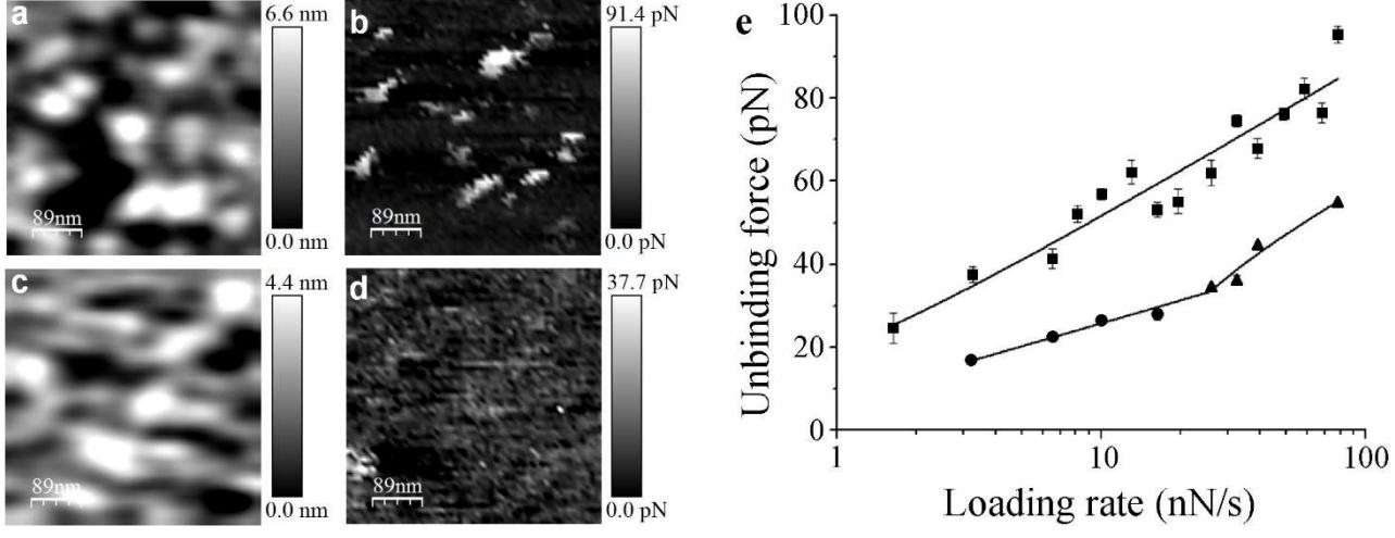
Atomic force microscopy (AFM) emerged as promising technique to address multitude of biological system properties at the single molecule level. This tool provides high-resolution imaging by scanning the surface of almost any type of sample with ultra-sharp tips. Moreover, AFM allows to acquire data under liquid environment mimicking the intracellular conditions inner the living cell or bacteria. This aspect becomes AFM more appealing in comparison with other scanning probe (SPMs) or electron microscopies (TEM) when the study is focused on biomolecular systems. Here, we overview the potential applications of AFM and their operational modes by real examples that our group has been deeply studied in the last decade. First, we present AFM as suitable technique to gather topography information with sub-nanometer resolution in aqueous media. Several examples are showed: I) The analysis of the FAD synthetase from Corynebacterium ammoniagenes upon ligand binding and catalysis [1] and II) The conformational study of the novel family of Ferredoxin-dependent Flavin Thioredoxin Reductases [2], both through imaging. Then, the methodology to immobilize in an oriented manner the Ferredoxin NADP+ Reductase (FNR) flavoenzyme on a substrate towards its redox partners Ferredoxin (Fd) and Flavodoxin (Fld) [3] and use this strategy for molecular recognition imaging (MRI) and do force spectroscopy (FS). MRI enabled us to identify biomolecules on mixtures immobilized on surfaces by unbinding force mapping. This approach reaches to quantitatively discriminate FNR molecules on a sample through the rupture of the aforementioned FNR:Fd complexes using Fd- functionalized tips (Fig. 1, a-d) [3]. Finally, the study of the intermolecular forces and energy landscapes through dissociation of complexes under force using dynamic FS between immobilized FNR and Fd and Fld molecules covalently linked onto the AFM tip [4]. The results indicated that the FNR:Fd complex is more specific, mechanically stronger and more durable than FNR:Fld. In addition, Fd dissociates from FNR through a single barrier while Fld followed two energy barriers (Fig. 1 e).