tBLMs that mimic biological cell membranes have become broadly used in studying lipid-protein interactions and the construction of biosensors for toxin detection. It is quite often used in synergy with vibrational spectroscopy techniques such as surface-enhanced Raman or infrared absorption spectroscopy in-situ (SERS and SEIRAS accordingly). Such systems can generate copious amounts of data providing information and complexity at the very same time. The incorporation of molecular markers can help to identify various physical and structural properties of the different domains in the system.
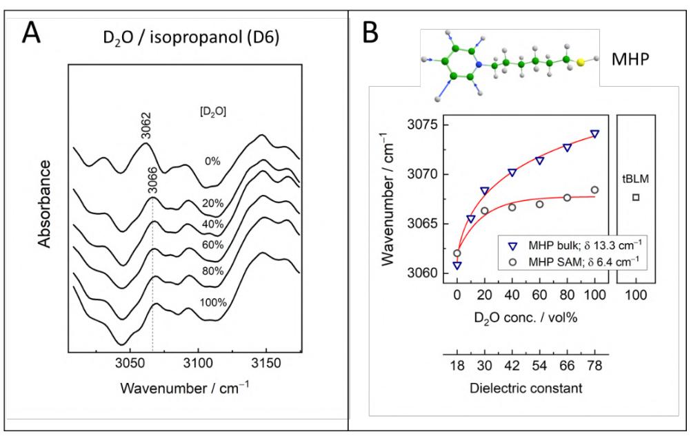
In this work, we used the MHP (N-(6-mercapto) hexylpyridinium, [1]), lipid-like molecule WC14 (20-tetradecyloxy-3,6,9,12,15,18,22-heptaoxahexatricontane-1-thiol), and βME-D4 (deuterated 2-mercaptoethanol) to form tBLM anchoring self-assembled monolayer (SAM). The MHP, spectral marker molecule, allowed us to probe the dielectric environment of the submembrane layer of tBLM. SAM formed on ~25 nm SEIRAS potentiometrically activated gold layer on silicon ATR prism [2] was monitored by FTIR spectrometer (Bruker, Germany). Spectral marker at 3062 cm-1 assigned to stretching =C–H vibrations of pyridinium ring monitored within a series of different ratio D2O/Isopropanol solutions from 0 to 100% (Fig. 1, A). Water addition to the solution has an impact showing absorption band shift by $\delta$ = 6.4 cm-1 towards higher wavenumbers. A similar experiment of MHP in solution with double wavenumber shift (13.3 cm-1) (Fig. 1, B) indicates lowered dielectric constant of the environment in the SAM layer. Which is explained by a less hydrophilic environment as MHP is surrounded by WC14 and βME-D4. Additionally, we determined an unchanged peak position value after the formation of tBLM membrane on SAM. It seems logical, that the additional membrane layer does not change the environment of MHP because lipids are located further from the surface.
Overall, MHP as a marker can be used to indicate the dielectric constant of the local environment. However, changes in the pyridinium ring stretching C–H vibrations become rather small after 20–30% D2O concentration has been reached. Additionally, the potential of MHP as a hydrophobicity marker of the gold-SAM interface has been revealed, which needs additional study.