Quantum dots (QDs) are nanoscale semiconductors, usually consisting of a crystalline core surrounded by a protective shell [1]. QDs are currently used in various fields of science, especially in biomedical applications such as drug delivery, gene therapy, bioimaging, etc. [2]. The core of conventional QDs contains potentially toxic substances (eg., Cd, Te), so less harmful elements, such as Cu, can be used as an alternative [3]. Many core-shell combinations and unique properties provide the plethora of QDs, so it is important to study them under natural environmental conditions, such as prolonged exposure to light, and media with various ions and biomolecules, to obtain information about their fate, after QDs are released into the environment.
In this work, we monitored the photostability of hydrophilic CuInS/ZnS-COOH QDs (Nanooptical materials, USA) in various biological media by measuring changes in optical density (OD) and photoluminescence (PL) spectra. After preparation of QDs samples (the main concentration was 4 nM), both OD and a peak PL intensity at 548 nm were similar in deionized water and in MWC algae growth medium. The peak position of the PL spectrum of QDs in deep well water remained the same, but the initial PL intensity was lower, and scattering was recorded in the spectrum of OD. During the first week, the spectral properties of QDs stored in the dark at room temperature (18 °C) practically did not change, regardless of the medium.
Some QDs samples were also stored at room temperature, but under continuous white light illumination (Osram Dulux Star, 11W/827). For another part of the samples kept in the Shrimp Set Smart aquarium (Aquael, Poland) at about +22 °C, an 8h/16h light/dark cycle was applied using a LED lamp (6 W) designed forgrowing aquatic plants. Regardless of the medium, both the OD and the PL intensity decreased in the illuminated samples, but this decrease was dose-dependent: samples that were under continuous white light illumination ceased to produce measurable fluorescence signals within three days, while samples exposed to light/dark cyclic illumination – after five days.
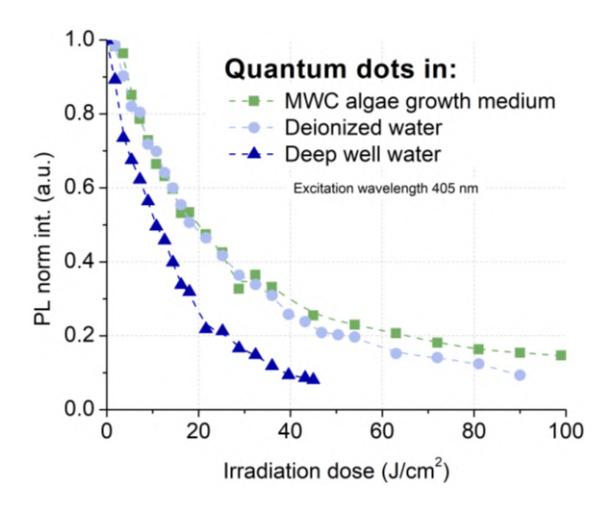
During the irradiation experiment, the QDs samples were prepared in a 12-well plate, and the collimated violet LED light (404 nm, 30 mW/cm2) was directed from below the plate from a distance of 8 cm. The fractionated exposures were delivered for 20 minutes to obtain the partial cumulative dose up to 36 J/cm2 (in intervals of 1.8 J/cm2 and 3.6 J/cm2) in all media, and from 36 J/cm2 to the total dose up to apparent decrease in the PL intensity (in intervals of 3.6 J/cm2 and 9 J/cm2) according to the samples. Initially, the PL intensity decreased in all media (in deep well water faster than in other), but after reaching approximately 21.6 J/cm2 (in deep well water) and 28.8 J/cm2 (in deionized water and MWC algae growth medium) the decline slowed down (Fig. 1). The spectral properties of CuInS/ZnS-COOH QDs can be considered so photo-unstable that the effect of the medium is negligible compared to light, despite the initial spectral differences.