
Saccharomyces cerevisiae yeast adapted to employ HOG pathway to recover after dangerous shape modifications and intracellular water disbalance caused by environmental osmotic pressure changes [1]. Pulsed electric field (PEF) treatment is known to cause plasma membrane permeabilization, an effect known as electroporation, yet at the moment there is no information on whether HOG biochemical pathway has a role in yeast response to PEF treatment [2].
It was investigated if post-PEF osmotic pressure change of the media has any effect on yeast cells' viability and size and if HOG pathway is involved in recovery. Experiments were performed with wild type Y00000 yeast and a mutant strain derived from WT, Y02724, with no active HOG1 gene. Cells were grown until 1OD, transferred to electroporation buffer (20 mM TRIS, 1 M sorbitol, pH = 7.4), yeast suspension was exposed to single electric field pulse with duration of 150 μs and field strength of up to 10 kV/cm. Electroporation buffer was used as a reference point to represent isoosmotic conditions. Swift osmotic pressure change is defined as osmotic shock. After PEF treatment cells were immediately transferred to hyperosmotic (1.5/2 M sorbitol), isoosmotic or hypoosmotic (0.1/0.5 M sorbitol) solution, incubated for 5 minutes and plated on solid YPD media. After two days of incubation in 30 °C CFU were counted. Cell size was calculated by measuring the turbidity of the solution after 10 seconds and 30 minutes of osmotic shock treatment [3].
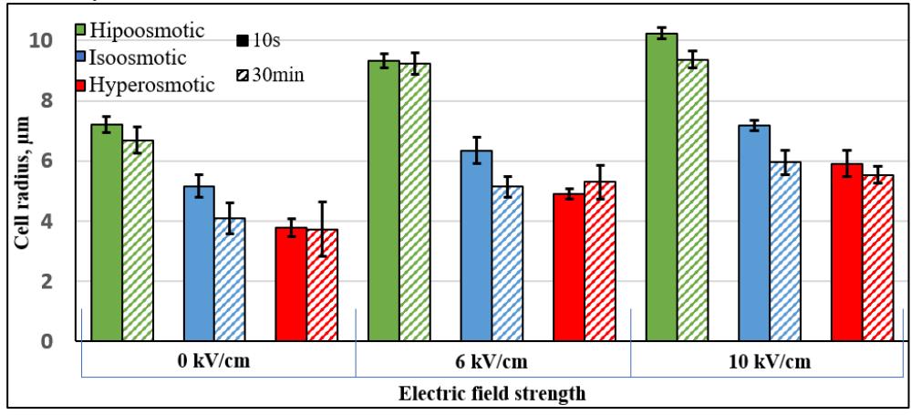
It was shown that post-pulsed electric field treatment by sudden change of osmolarity of the media has a significant impact on the yeasts' viability, cell size and leakage of intacellular components into the media when compared to isoosmotic conditions. Thus we show that electroporation efficiency is not only dependent on electric field strength, duration and the number of impulses applied, but also on whether post-PEF treatment was applied. While it is known that electric fields of 8-10 kV/cm causes irreversible damage to membrane, after incubation in hyperosmotic conditions after PEF viability increased by ~20-30% and radius of the cell was reduced by as much as ~2 μm (Fig.1) After weaker field strengths of 2 and 4kV/cm PEF and hyperosmotic shock treatment viability of WT yeasts was restored to 100%, for ∆hog such result was achieved only after 2 kV/cm treatment. Hipoosmotic shock, evidently, caused the opposite effect: after 4 kV/cm viability decreased by 12% (WT) and 39% (∆hog); after 6 kV/cm by 16% and 25%. The size of the cells increased almost two times, after 10 kV/cm field (Fig.1)
Protein and thiol group concentration evaluation revealed that amount of intracellular compounds in the media decreased after hyperosmotic shock and increased after hipoosmotic shock, supporting the hypothesis that mechanical cell shape alteration influences cells' reaction to PEF. Furthermore, ∆hog strain was more sensitive to beforementioned treatments suggesting that HOG pathway is of relevance to recovery after electroporation.
To summarise, yeast cell viability and size after exposure to PEF can be altered by subsequent change in osmolarity of media. HOG pathway involvement was linked to recovery after electroporation.