Liver cancer is one of the most frequently diagnosed malignancies and is one of the leading causes of cancer related deaths worldwide [1]. Malignant neoplasms in liver could form as primary liver tumors or as metastasis from other organs. In both cases, partial hepatectomy is the standard treatment for liver tumors. During the surgery, the tumor and a small amount of the surrounding normal tissue are removed to ensure the complete resection. The success of the treatment depends on the ability to determine the exact border between normal and malignant tissues. All the malignant tissue should be removed in order to prevent the recurrence of the tumor. On the other hand, it is important to preserve as much as possible of normal tissue to maintain functions of the organ. Therefore, a method that could be used directly in the operating room during the surgery and could give a reliable and fast diagnostic result, is needed.
In recent decades, a variety of methods of vibrational spectroscopy are applied for the examination of biological tissues [2]. Using ATR (attenuated total reflection) technique, samples can be examined without any specific preparation. In this study, the fiber ATR IR probe was applied for the spectroscopic measurements that allows to investigate samples ex vivo under native conditions what is important during the surgery.
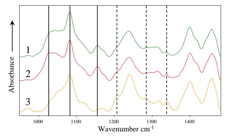
In this study, 25 cases of patients with liver tumors were investigated. Measurements were performed for two types of samples: (I) the tissue immediately after the surgical removal (13 patients) and (II) frozen tissue (12 patients) samples. In both cases normal and tumorous tissues for each patient were investigated. Samples of freshly resected tumorous tissue included primary tumors of liver (hepatocellular carcinoma (HCC), cholangiocarcinoma (CCC)) and metastasis from other organs (colorectal carcinoma (CRC), pancreatic adenocarcinoma (PDAC) and oesophagogastric junctional adenocarcinoma (EGJA)). Samples of frozen tumorous tissue included primary liver tumors (HCC and CCC). The spectra of normal, HCC and CCC liver tissue are represented in fig. 1. Spectral bands of higher intensity located at 1028, 1082 and 1155 cm-1 can be attributed to glycogen and they are observed in spectra of normal (1) and HCC (2) liver tissue. Such findings can be explained considering liver function accumulating glycogen in hepatocytes. Intensity of the glycogen spectral bands decreases in the spectrum of the CCC tissue. Whereas CCC tumors form out of epithelial cells of the biliary tract, level of glycogen in CCC tumorous tissue is reduced compared to normal liver spectra. Besides the decreased intensity of the glycogen spectral bands, increased intensity of collagen spectral bands located at 1207, 1283 and 1338 cm-1 is clearly visible. The increased intensity of these spectral bands indicates higher amount of collagen in the tissue. Increased level of collagen is related to desmoplastic stroma of the tumor that is rich in connective tissue containing collagen and is characteristic for CCC tumors. In case of HCC tumor, spectral differences between normal and tumorous tissue spectra are not clearly visible. Despite of this fact, after application of hierarchical cluster analysis (HCA), spectra of normal tissue can be classified with 88 % accuracy while spectra of tumorous tissue (including HCC tumors) - with 80 % accuracy.