For the last few years, the attention and interest in metal nanoparticles, their applications, and possible synthesis methods is noticeably increasing faster than ever before. This is mainly because of the specific and unique nanoparticle's chemical, optical, electric properties, compared with their bulk counterparts. Additionally, nanoparticles can be used for numerous practical applications, like catalysis, medicine, biotechnology, and so on [1]. Different synthesis methods of nanoparticles exist, but laser ablation in liquid (LAL) method seems to be one of the most favorable because the system itself is simple and it allows to produce nanoparticles of small size range deviation, varies their morphology, and produced nanoparticles are contamination-free when no additional surfactants are used in the process [1].
The purpose of this work was to produce silver nanoparticles using LAL of a silver target (99.9%, JSC Lithuanian Mint). The influence of the ablation media (ultrapure type 1 water, chloride, and iodide salt solutions) was related to the size and shape of the resulting nanoparticles. The laser ablation was carried out employing a first harmonic of 1030 nm of a 270-fs pulse length Yb:KGW femtosecond laser (Pharos, Light Conversion) operating at a 200 kHz repetition rate. A galvoscanner and a telecentric f-Theta lens equipped in a laser micromachining workstation (FemtoLab, Altechna R&D) were used. The analysis of the shape and size of produced silver nanoparticles was performed using scanning electron microscopy (SEM, Quanta 200 FEG, FEI) and software Ilastik whereas the optical properties were evaluated using UV – vis spectroscopy (AvaSpec 2048, Avantes).
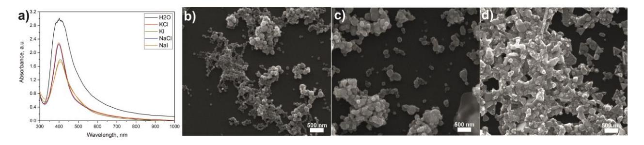
The successful formation of the silver colloidal solutions was confirmed by their unique distinguished yellow color. Absorption spectra of silver colloidal solutions synthesized in different environments are depicted in figure 1, a. The absorption peak corresponds to localized surface plasmon resonance of silver nanostructures [2] and varies from 398 nm to 408 nm depending on the ablation media.
Even though, using different media for target immersion the optical spectra did not change considerably, the SEM analysis revealed that it had an expressed effect on the shape and size of nanoparticles as is seen in Figure 1, b-d. From the results, it was observed that halogen etching and photo-irradiation using laser beam leads to the conversion of silver nanoparticles morphology to more complex geometries than spheres observed in the case of water [2]. The diameter of spherical silver nanoparticles produced in ultrapure water was in the range of 15 – 25 nm. On the other hand, nanoparticles produced in chlorides and iodides solutions demonstrate nanotriangles, nanocubes, and nanorods morphologies. Length of the edges of such nanostructures vary from 100 nm to 400 nm. Obtained results show that changing the immersion liquid LAL synthesis of metal nanoparticles can be effectively used to produce silver nanoparticles of different morphology.