
One of the first 3D printing technologies is stereolithography (SLA), which creates structures by selectively solidifying a liquid resin during photopolymerization reactions and has attracted considerable interest in electronics, biomedicine [1], and more over the past decade. In this work, the application of SLA printer in tissue engineering was investigated. It is an interdisciplinary branch of science that involves the use of complex scaffolds and cells to create functional tissues that would replace damaged tissue or missing organ. This treatment can overcome the obstacles of traditional methods, such as the lack of organ donors and the rejection of transplanted tissues. One of the main problems in printing scaffolds for tissue engineering is the selection of biocompatible materials, biomimicking geometries, and post-exposure processing[2].
The aim of this report is to present the fabrication of 3D micro-porous scaffolds from commercial resins and their application to cell growth. The study was performed using $Asiga\ PICO2$ device, finding the optimal printing parameters for $Formlabs\ Clear$ and $Formlabs\ Flexible$ resins. The critical time $T_k$ and the penetration depth $h_a$ were calculated according to Beer-Lambert's law as proposed earlier [3]. Based on the obtained parameters, a study of printer evaluation to create samples of different geometries for two materials, was performed. The qualitative parameter (A) of repeatability of the printed structures was calculated on the basis of it samples with different thickness gaps and building blocks, different materials and geometry were compared. Quantitative biocompability MTT studies [4] supported by qualitative scanning electron microscope (SEM) and fluorescence microscope studies were performed after printed scaffolds were processed with different post-exposure methods to investigate biocompatibility and cell morphology.
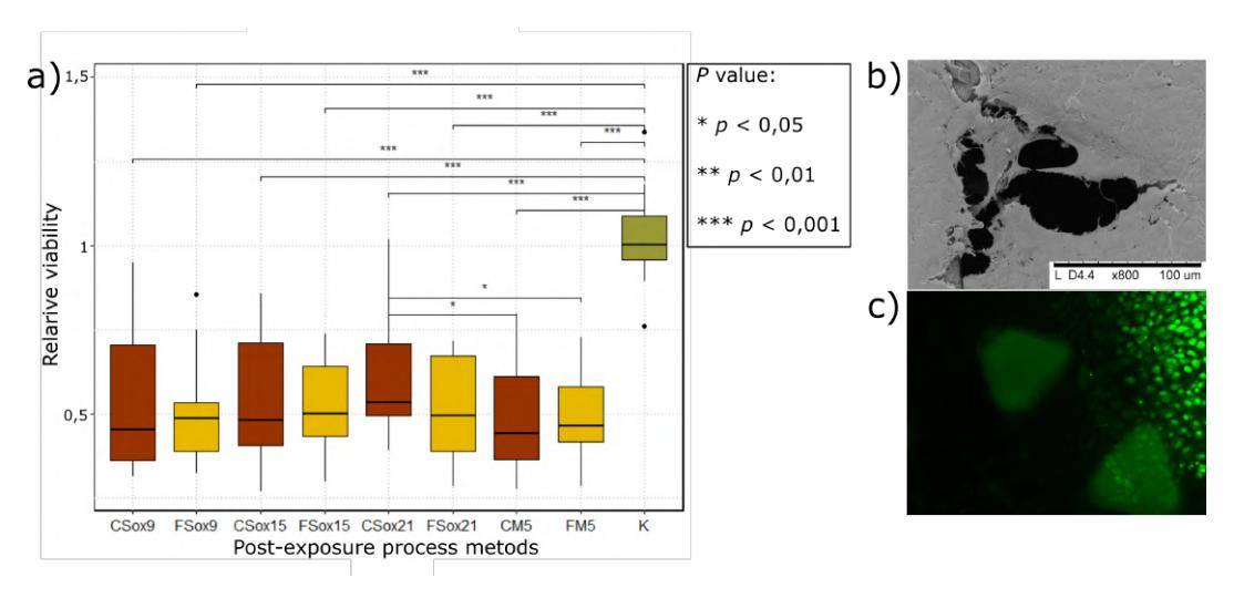
It was observed that it is more difficult to print rare geometry (fine strings and broad gaps) materials with Formlabs Clear resin than with Formlabs Flexible. The investigated repeatability parameter A shows that by changing the geometry of the structure (in order: rectangle, square, triangle), obtained values were decreasing. The assessed biocompatibility of differently treated scaffolds showed that none of methods completely removed the monomers but all were suitable for cell growth. The materials showed similar results with each other. A superior performance was observed in the sample soaked in methanol and the Soxhlet extractor using ethanol. Quantitative MTT biocompability results shown in Fig. 1. Also, validated by a qualitative biocompatibility study and depicted cell arrangement on 3D scaffold.