
Approximately 5.2% of the elderly population worldwide suffers from some form of dementia, of which Alzheimer's disease (AD) is the most common cause. Currently there is no definite diagnostic test for AD, other than post-mortem examination. However, one of the early symptoms of AD is the impairment of visual processing. Studies show that the amplitudes of visual evoked potentials (VEPs) are reduced in AD and amnestic mild cognitive impairment (aMCI), an interim stage between healthy aging and dementia. (Fig.1) [1]. VEPs are peaks in electrical potentials within neural populations as a response to a visual stimulus, measured using an Electroencephalogram (EEG). Mathematical modelling can be used to investigate neural mechanisms causing reduced VEPs, and identify biological parameters that could be biomarkers of AD in the brain. In this work, we fitted a neural mass model [2] to clinical VEP data [1] using an evolutionary algorithm. We analysed subject-specific sets of model parameters to understand the effect of AD on the neural architecture and dynamics of the visual pathway.
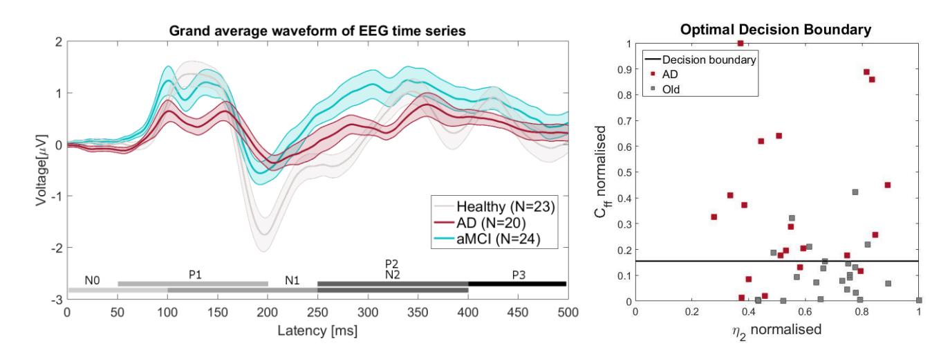
We found that self-inhibition of fast-spiking inhibitory interneuron population (FS) is increased in AD and aMCI compared to healthy aging subjects, whereas the inhibition of pyramidal population by FS cells is reduced in aMCI and AD. Based on the self-inhibition parameter (Cff), we classified subjects into healthy and AD on the one hand (Fig.2) and healthy and aMCI conditions on the other with an accuracy of 76.7% and 72.3% respectively. Our results are in line with previous findings, whereby impaired inhibitory function of FS cells has been linked to deficits in information processing, indicating the potential for AD drugs to target inhibitory neuron populations. The success of classification suggests that parameter-based VEP analysis could provide a novel non-invasive tool in the diagnosis of aMCI and AD.