
The usage of biosensing systems is promising - rapid and accurate method for detection and analysis of various compounds. Nowadays one of the best-known and the most common biosensors are the glucose biosensors. They can accurately detect concentration of glucose in blood during a short period of time – it is very important to people with diabetes [1]. The active part of the biosensor is a glucose oxidase (GOx) enzyme immobilized on the surface of the electrode. When constructing an enzymatic biosensor, one of the most important aims is to determine properties of an enzyme under different environmental conditions. Despite the increasing number of studies on GOx, there is little information about the properties of the enzyme at different pH conditions [2].
Spectroscopic properties of glucose oxidase (GOx) and flavin adenine dinucleotide (FAD) were investigated in different acidity environments. The purpose of this research was to evaluate absorption and fluorescence spectra changes. The study of the absorption and fluorescence spectra and the measurements of relaxation times were carried out using a buffer with pH values from 2 to 8.
GOx is a flavin adenine dinucleotide (FAD) containing glycoprotein. FAD is involved in enzymatic redox reactions and determines the enzyme's activity [3]. Glucose oxidase enzyme can exist in several different spatial conformations, therefore, FAD can also assume “twisted” (stacked) or “planar” (open) structure [4].
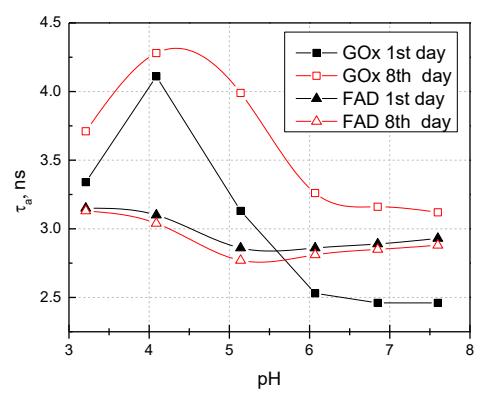
Fluorescence lifetimes of GOx and FAD have been measured at fixed 530 nm wavelength. The fluorescence decay kinetics were well approximated by two exponents, those were used to calculate the average relaxation time. Fig.1. shows the changes of average fluorescence lifetimes at different acidity of a buffer in the 1st and 8th days. The short component of fluorescence decay is associated with the "twisted" structure of FAD and the long component is associated with the "planar" spatial configuration of FAD. In a planar configuration, intramolecular electron transfer between the flavin isoalloxazine ring and the adenine moiety takes a longer time.
The data analysis showed that long component of FAD's and GOx's fluorescence decay dominated in the acidic environments. In alkaline solutions of FAD, the ratio of a short and long component begins to change from 1:16 to 2:3. The alkylation of GOx solution also starts exchanges of a ratio of short and long component, but it is not as significant as in the FAD's case.
During this study, it was found that at pH 3 solution acidity, the fluorescence intensities of FAD and GOx at 530 nm were the most intense. At the optimum pH (6) fluorescence intensity of GOx was the lowest. The increased intensity of the fluorescence band of GOx is associated with dissociation of FAD from the enzyme. Also, the increase of GOx fluorescence is due to the substantially increased fluorescence of FAD at the acid buffers (pH 3 - 4).