
The rapid technological development in the medical field has led to implementation of new vibrational spectroscopy methods for research on molecular biology. It was shown that these methods can be used to identify the cells of kidney, breast, and prostate cancer [1].
An overall success of removal of cancerous kidney tissue during the surgery is highly dependent on fast and accurate scoping of kidney tissue. Unfortunately, clinical methods used nowadays cannot ensure this due to time consuming preparation of the samples. Thus a demand for new procedures is growing.
It is known that cancerous and healthy kidney cells differ in metabolism and molecular structure [2]. For this reason, the extracellular fluid of cancerous and healthy kidney tissues also differ since the extracellular fluid composition is closely related to the metabolism of the cells. Thus, due to a simple preparation of the extracellular fluid samples, a faster identification of cancerous cells can be achieved by analyzing the collected vibrational spectra of extracellular fluid. However, conventional Raman scattering cannot be effectively used for thin film analysis and unconventional methods like surface enhanced Raman scattering spectroscopy must be used.
Since biological fluids are composed of different molecules, their experimental spectrum is a combination of many different overlapping vibrational spectral bands. Thus, precise assignment of these bands to the corresponding molecular vibrations is a difficult task. The intensity of SERS spectral bands depends on the distance between the molecule and a metal surface, which is used for the SERS effect. When the samples are cooled, the distance between molecules and the nanoparticles should decrease and the enhancement factor of the SERS signal of the adsorbed molecules should increase. When the sample is composed of different molecules, the adsorption also depends on the functional groups of the molecules in the composition of the sample.
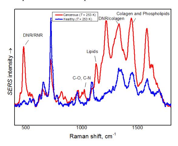
In this study, low temperature SERS measurements of extracellular fluid of cancerous and healthy kidney tissue samples were carried out in order to get a deeper understanding of the nature of vibrational spectral bands present in the experimental spectrum.