
In vivo, cells are embedded in a 3D microenvironment which influences their behaviour and signalling. To mimic that environment, cells can be cultured on 3D scaffolds in vitro. There are currently over 40 different 3D printing technologies that can be used for this purpose. Stereolithography is one of them and it may be appealing for the fabrication of customised scaffolds with controlled pore size. Such scaffolds can be applied for tissue engineering purposes or used to study cell-matrix interactions as well as intracellular signalling [1, 2].
The main goal in this research was to test the capabilities of 3D printer Formlabs Form1+ and the appropriateness of Formlabs Flexible and Formlabs Clear materials for elastic scaffolds and tissue engineering.
First, the optimal printing parameters were determined by fabricating objects under varying conditions and scaffold porosities (Fig. 1). Then, primary rabbit muscle cells were seeded on the scaffolds and their proliferative activity was measured using a colorimetric assay based on the reduction of a tetrazolium salt. The experimental scheme is depicted in Fig. 2.
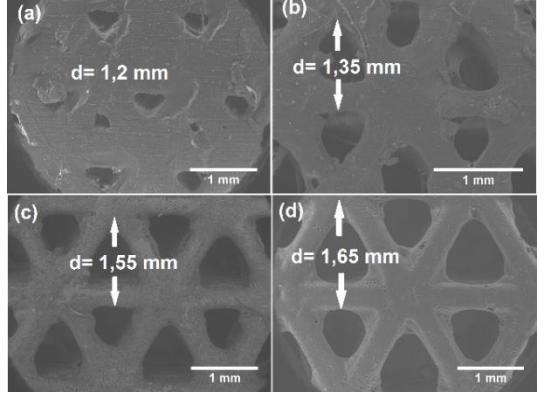
We found that scaffold layers tend to overlap in structures that are less than 3 mm tall, resulting in 2D structures. Biocompatibility tests showed that both the materials reduced cellular viability. We hypothesised that the source of such toxicity were uncrosslinked monomers within the material, so additional UV treatment was investigated as a possible solution to the problem. However, our results did not reveal cell viability correlation with UV curing time.
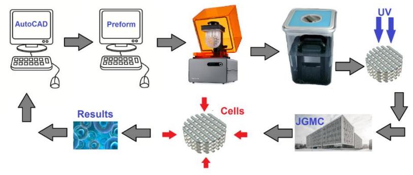
A 3D printer with better control over polymerisation conditions might help improve the biocompatibility of printed scaffolds, so we are currently testing Autodesk Ember 3D printer, which has a higher resolution and provides more tuneable printing parameters.