
Positron Emission Tomography (PET) and Single-Photon Emission Computed Tomography (SPECT) are 3D nuclear medical imaging techniques which use radiopharmaceuticals administered to the patient and a gamma scintillation camera to produce functional images of metabolic activity in the patient's body. The radiopharmaceuticals emit gamma photons through radioactive decay (either directly in SPECT [1] or through positron-electron annihilation in PET [2]) and the signal is registered by a gamma scintillation camera, which is rotated around the patient's body to provide a set of projections, which in turn are processed using tomographic algorithms to create a 3D model of the distribution of the radiopharmaceutical in the patient's body. As the radiopharmaceutical is selected to accumulate in areas exhibiting metabolic activity of diagnostic interest, these techniques permit for noninvasive mapping of these types of metabolic activity.
Of particular interest for this research is the application of SPECT for imaging the heart. However, both PET and SPECT are prone to a few types of interference. Despite being selective, the radiopharmaceuticals used in these techniques accumulate in other organs in addition to the heart (particularly in the liver). In certain projections, the signal from those organs is difficult to distinguish from the signal from the heart. Moreover, due to their anatomy, the lungs have a non-uniform coefficient of attenuation of the gamma signal [3]. For these reasons, even the most advanced computationally-intensive iterative reconstruction algorithms used to stitch the individual projections into a 3D model have trouble providing precise images.
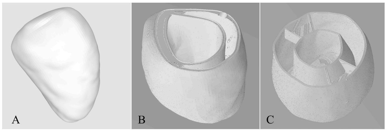
In order to improve PET and SPECT imaging technology, an accurate anthropomorphic phantom of the human chest is necessary so that modifications to the equipment and the processing algorithms can be tested in controlled settings. Commercially available phantoms suffer from several limitations, the most important ones being low detail, cost, and usability. We set out to create a highly accurate set of phantoms of the heart, representing both a healthy heart and hearts with various defects. Due to the internal complexity of the model, 3D printing was the obvious choice for the manufacturing technique. One of the challenges in 3D printing such a phantom is the fact that it must be watertight and reusable so that it may be repeatedly filled with solutions of the radiopharmaceuticals used in PET and SPECT. Working with a team of 3D design and 3D printing experts, we explored various materials, printing techniques, and processing techniques to produce these phantoms.