
An important problem in cell technologies is to develop the methods of accumulation of cells with certain characteristics [1, 2]. The aim of this study was to choose the electrical stimulation modes which can provide a changing the cell proliferative activity. Epithelioid tumor cells of human cervix carcinoma (line HeLa) were used as object of the research. Cells were cultivated in DMEM 1152 supplemented with 25 mmol/L HEPES, 10% FBS at 37 °C and 5% CO2 content. Cell culture was stimulated by uniform periodic pulsed electric field using programmable electrical biological stimulator (BSU, Minsk, Belarus) with platinum electrodes. Electric stimulation modes differ in number of pulses in series (1, 3 or 5) and amplitude of the pulses (6.6 or 20 V/m). The frequency of pulse series was 10 Hz. The stimulation lasted for 12 h on the first or the second days after passaging.
It was revealed that in the absence of electric stimulation the cell doubling time of HeLa cells was 21-23 h. The stimulation of HeLa cells in the first culture growth day (from 8 to 20 h after passaging) led to significant amplification of cell proliferation at pulsed electric field intensity of 20 V/m (fig.1 A, modes 1 and 2). When the electric stimulation was carrying out on the second day of culturing (from 24 to 36 h after cell passaging) the proliferation enhancing effect was reduced (fig.1 B, modes 1 and 2). The changes in proliferative activity of HeLa cells were insignificant at lower electric pulse amplitude of 6.6 V/m under stimulation both the first and the second days (fig. 1 A and B, mode 3).
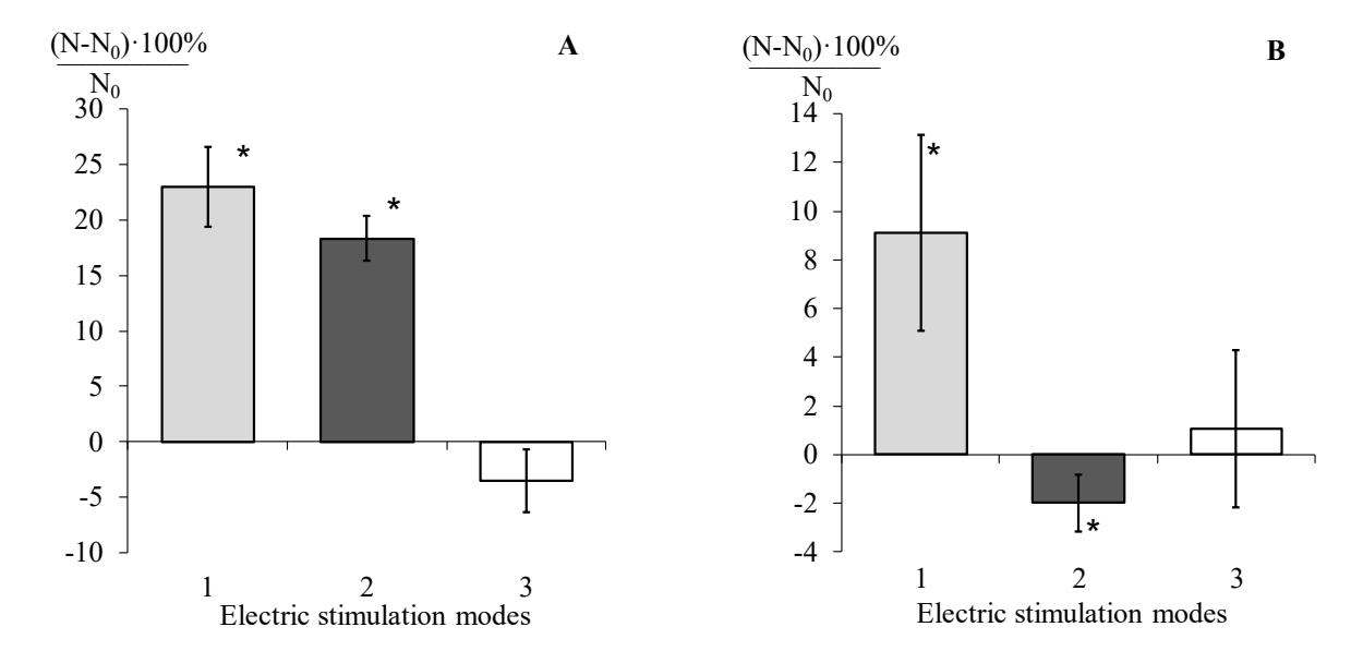
The mechanism of electric field action on cells can include changes in ion channels activity, values of transmembrane electric potential, cell surface charge and as a consequence modulation of cell ability to adhesion and division [1-3]. The obtained data confirm the possibility of regulating cell proliferative activity by exposure to a periodic pulsed electric field. In particularly, HeLa cell proliferation is amplified under influence 20 V/m electric pulses with 10 Hz frequency during the first day of cultured growth.