
Cancer is the second leading cause of death (after heart diseases) in developed countries. Early and accurate detection of cancer using X-ray is valid, but complicated due to poor contrast of tissues. In order to enhance visibility of structures in body contrast agents were created. Water-soluble contrast agents made from iodinated molecules are clinically approved and widely used for X-ray imaging although sometimes can be insufficient. Different contrast agents were created and examined in order to overcome some limitations of iodinated contrast materials. Gold nanoparticles are considered to be one of the most reliable metal-based contrast agents which are expected to significantly improve X-ray imaging of soft tissues in vivo [1]. Gold nanoparticles (AuNP) have attracted intense interest as contrast agent over the past few years due to suitable properties for effective contrast material.
The aim of this study is to verify the best conditions of formation of polyvinylpyrrolidone (PVP) stabilised gold nanoparticles as well as to assess size distribution concentration and stability of AuNP dependence on reagents.
25 different synthesis products of AuNP were made by varying the concentrations of MES monohydrate and sodium hydroxide (NaOH) reagents. Synthesis was made at room temperature, by mixing MES and NaOH solutions, adding polyvinylpyrrolidone (PVPK30) and chloroauric acid (HAuCl4) and vigorously stirring for 1 hour.
To determine properties and concentration of produced gold nanoparticles absorption spectra were measured (Fig. 1). The further stability and processes were monitored by measuring absorption spectra after 1, 2, 10 and 30 weeks.
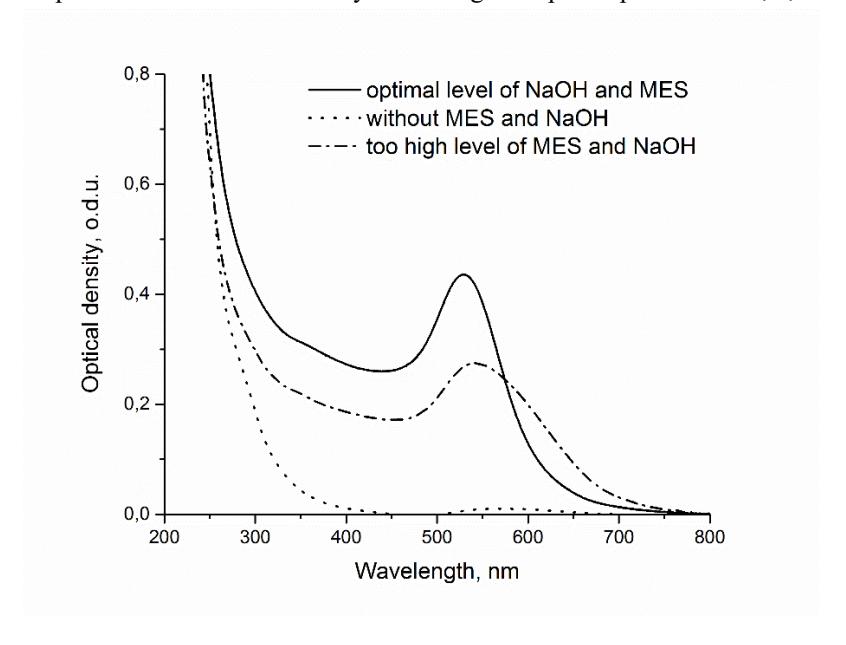
According to the absorption spectra (Fig. 1) the amount and size of produced AuNP depend on both concentration of reducing agent and pH. The sample without MES and NaOH had no significant absorption in comparison with other samples showing that synthesis did not occur. Nevertheless, when NaOH were added the absorbance appeared affirming that AuNP were produced. When NaOH and MES concentrations in primary solutions were optimal the concentration of uniformly sized nanoparticles was highest [2]. Although when concentrations of MES and NaOH exceeded these levels, synthesis of AuNP became not so efficient with higher distribution in size and decreased amount of formatted nanoparticles.
Results showed that concentration of reagents is critical for efficient synthesis of gold nanoparticles. Further investigation in conditions of AuNP synthesis will provide a more successful development of gold-based contrast agents and improvement in X-ray imaging.