
Photodynamic therapy (PDT) efficiency primarily depends on light source and photosensitizer (PS) properties. Many conventional PSs are hydrophobic and have limited tissue distribution. The upgrading of conventional PSs by the chemical modification alters the drug pharmacokinetics and biodistribution in tumor tissues.
The aim of this study was to assess the distribution processes of chlorin type PSs in human adenocarcinoma HT29 monolayer and spheroid cell cultures. We used the second generation PS chlorin e6 (Ce6) which is already accepted for clinical use and its dimethyl ester (DME) (Minsk, Belarus) [1]. It was shown, that the difference in chemical structure didn't significantly influence spectroscopic characteristics of PS, while some physicochemical properties such as water solubility were changed.
In the present work we compared the interaction of Ce6 and DME with HT29 monolayer cells. DME accumulated faster than Ce6 with the 2.3 times increased equilibrium uptake level. Moreover, it was demonstrated, that the chemical modification of Ce6 molecules leads to the changes of the PS intracellular localization and enhanced photosensitizing activity. Indeed, two times higher photokilling ability was registered for DME-based PDT compared with that of Ce6.
Ce6 and DME biodistribution processes in tumor tissue were studied on multicellular tumor spheroids model. The PS concentration in spheroid cells treated for 6 hours was estimated by flow cytometry and fluorescence microscopy techniques. It was shown, that DME was accumulated better and more homogeneous in HT29 spheroids, while Ce6 had a heterogeneous distribution with bright spots on the surface of spheroids (Fig. 1).
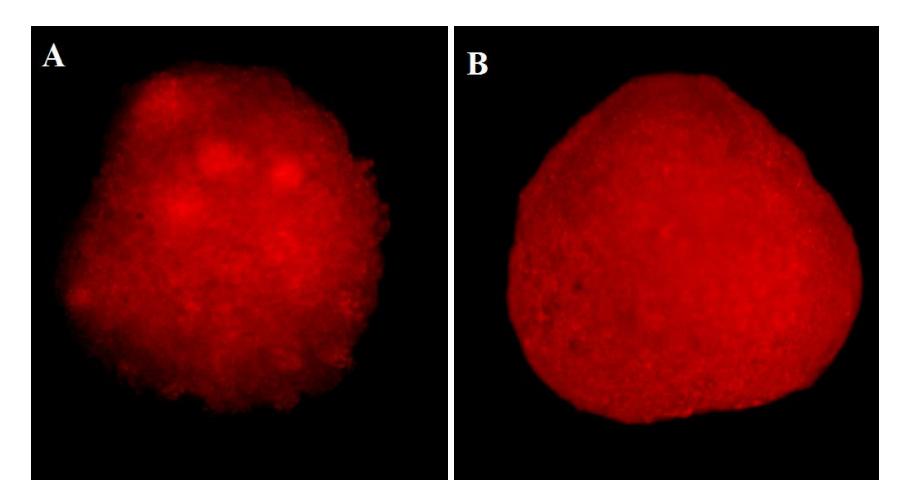
We suggest that improved accumulation, distribution and PDT efficacy of DME could be related to increased PS amphiphilicity and faster diffusion rate of DME molecules across biological membranes. Taken as a whole, we conclude that new chemically modified PS, based on Ce6, could be more efficient for the PDT of cancer, compared with conventional Ce6.