Anti-cancer treatment has become the vital point in medicine. Thus development of nanotechnology is one of the alternative ways of therapy. Iron oxide-based nanoparticles have been widely used for examining and treating cancer cells due to their magnetic properties in hyperthermia therapy.
Fe3O4 magnetic nanoparticles were synthesized in two different ways. Morphology and characterization of nanoparticles were measured with X-ray diffraction method, Scanning Electron Microscope and Superconducting Quantum Interference Device. First synthesis of Fe3O4 is based on high temperature decomposition of the self-synthesized Fe(III)-oleate. Different concentration of Fe(III)-oleate and time of the reaction resulted in various size of each nanoparticles. In second synthesis the magnetic particles were obtained by chemical precipitation from ferric (FeCl3) and ferrous salts (FeSO4) in alkali medium (ammonia hydroxide). To increase magnetic properties nanoparticles were doped with Yttrium in several concentrations.
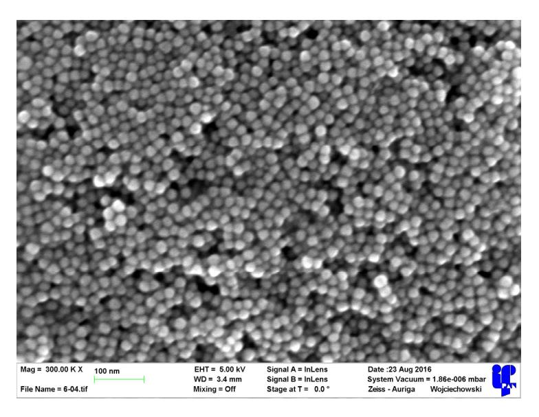
X-ray diffraction method confirmed a cubic single phase of Fe3O4 in both syntheses. SEM images showed different average size of nanoparticles from 10 nm to 25 nm. Nanoparticles from Fe(III)-oleate reaction had distribution of size around 2 nm and those obtained from precipitation around 6 nm. Measurements of relation between applied magnetic field (H) (Oe) and magnetization (M)(emu/g) varied from 9 to 84 (emu/g)(at 2000 Oe), where maximum value is obtained by doped Fe3O4. In 200 Oe field precipitated nanoparticles reached higher magnetization values quicker than those from Fe(III)-oleate, which went in favour of these nanoparticles obtained in the second reaction.
Measurements of influence of changing magnetic field on nanoparticles showed their ability to generate heat, which may be used in hyperthermia therapy.