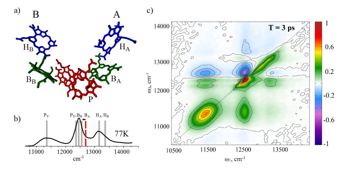
Reaction centers (RCs) from photosynthetic purple bacteria are pigment-protein complexes responsible for initial charge separation steps of photosynthesis. Following the isolation of RCs from photosynthetic bacteria, done in the late sixties, both arrangement of chromophores and the approximate structure of energy levels were determined. Nevertheless, due to the simple structure as well as high energy and charge transfer efficiency, which are not fully understood, the RCs is still one of the most investigated photosynthetic systems in the world [1].
The structure of quinones-depleted RCs (QDRCs) from purple non-sulfur bacteria Rhodobacter sphaeroides has six co-factors: two bacteriochlorophyll a molecules forming a strongly excitonically coupled dimer — a special pair (P), two monomeric bacteriochlorophyll a molecules (BA and BB) on each side of the dimer and two bacteriopheophytin a molecules (HA and HB). All pigments are arranged into two branches "A" and "B" featuring C2 symmetry (see Fig. 1, (a)) [2]. Remarkably, although excitations are absorbed and transferred to the special pair through both branches, the electron is transferred just through the active "A" branch [3]. After the charge separation in the special pair and formation of the P+HA- state, the monomeric bacteriochlorophyll molecule BA undergoes the electrochromic blue shift (indicated by an arrow in Fig. 1, (b)), which results in a well-known derivative shape signal, observed in the pump-probe measurements [4].
In this study, the QDRCs were investigated by the broadband two dimensional electronic spectroscopy at cryogenic temperature (77K). The study revealed dynamics with three characteristic lifetimes, which were observed also in the earlier studies: energy transfer of 100 fs, charge separation and electron transfer during 1.7 ps and a long (> 1 ns) lifetime for charge recombination. We also recorded the signals in the 2D spectra, arising from the band-shift phenomena and characterized them with the help of the double-sided Feynman diagram analysis. In addition, we observed the detailed features of the initial charge separation process, which have not been observed before.