
Ovarian and colo-rectal cancers are two of the most lethal malignancies, especially because of their natural progression from organ localized tumor to disseminated cancerous nodules around the whole abdominal cavity, namely peritoneal carcinomatosis [1].
Previously, this latest stage of cancer progression was considered as incurable. However, determined effort of surgeon, and practical approach of this pathology has led to the development of advanced surgical procedure. Nowadays, an effective cure, with high survival rates and acceptable life quality, is possible now by associating extensive surgery - to excise all tumor nodules, with advanced chemotherapy – to achieve residual cancerous cells.
Nevertheless, this achievement is still an awkward surgical challenge: if only one tumor, even the smallest, can escape from the surgeon's attention, survival rate is immediately decreased in two or more times, depending on the tumor size [2]. Moreover, surgeons still need effective techniques to detect tumors, in association with their only views and sense of touch.
To fulfill this request, fluorescence guided surgery (FGS) seems to be one of the most promising innovative technology. By using fluorescent dyes to stain cancerous tissues, and specific light sources and cameras, some research teams have improved the detection of tumors nodules, and even made visible the previously invisible smallest implants. These first steps of FGS are paving the way to better cure and easiest surgical operation [3]. However, many challenge remains, before using of this technology on humans.
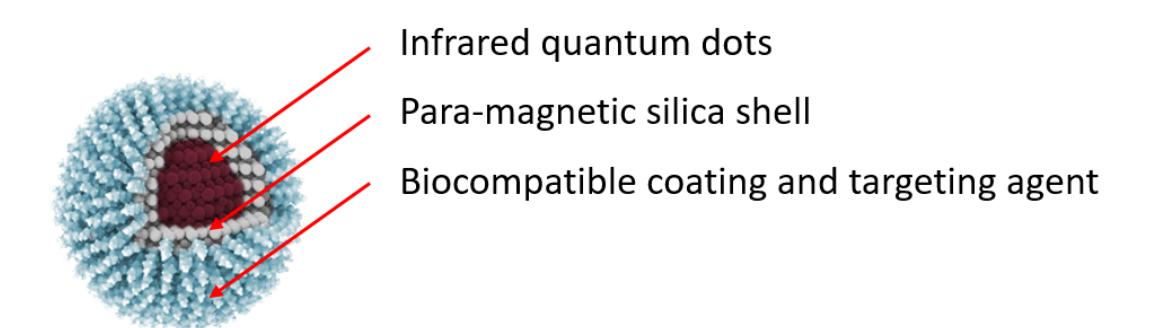
In the present work, we used the quantum dot based nanoparticles for ovarian peritoneal carcinomatosis imaging by magnetic resonance imagery, and to guide surgery by fluorescence. For this purpose, we conceive and upgrade an advanced kind of nanoprobe for guided surgery: by associating the exceptional fluorescent properties of quantum dots, in a biocompatible shell of mesoporous silica, doped with magnetic particles, we are developing an effective multimodal (i.e. fluorescent and magnetic) nanoprobes (Fig. 1).
After successful assessment of biocompatibility in vitro, we are now focused on chemical improvement to enhance in vivo biocompatibility of the probe. Also, we are developing murine model of ovarian peritoneal carcinomatosis, to assess imaging capability of our nanoparticles.