
Environmental pollution is currently being raised by a rapid application of nanoparticles in various fields. However, toxicity properties of nanoparticles are little-known. Embryonic development is an important model for studying the toxicity, in vivo system damage, transport and biocompatibility of nanoparticles and drugs.
The aim of this study is to examine distribution of CdSe/ZnS-COOH quantum dots (QDs) in rainbow trout (Oncorhynchus mykiss) embryos chorion using confocal microscopy and 3D mathematical modeling.
Analysis was based on 3D imaging of autofluorescence signal of embryo chorion and CdSe, covered with an additional semiconductor layer (ZnS), carboxylate functionalized, negatively charged, water soluble QDs (CdSe/ZnS-COOH) photoluminescence using Nikon ECLIPSE TE2000-U confocal – fluorescence laser scanning microscope along with 3D reconstruction software (Fig. 1. A). QDs distribution in rainbow trout embryos chorion was estimated. The laboratory treatment was carried out in climatic camera under static conditions.
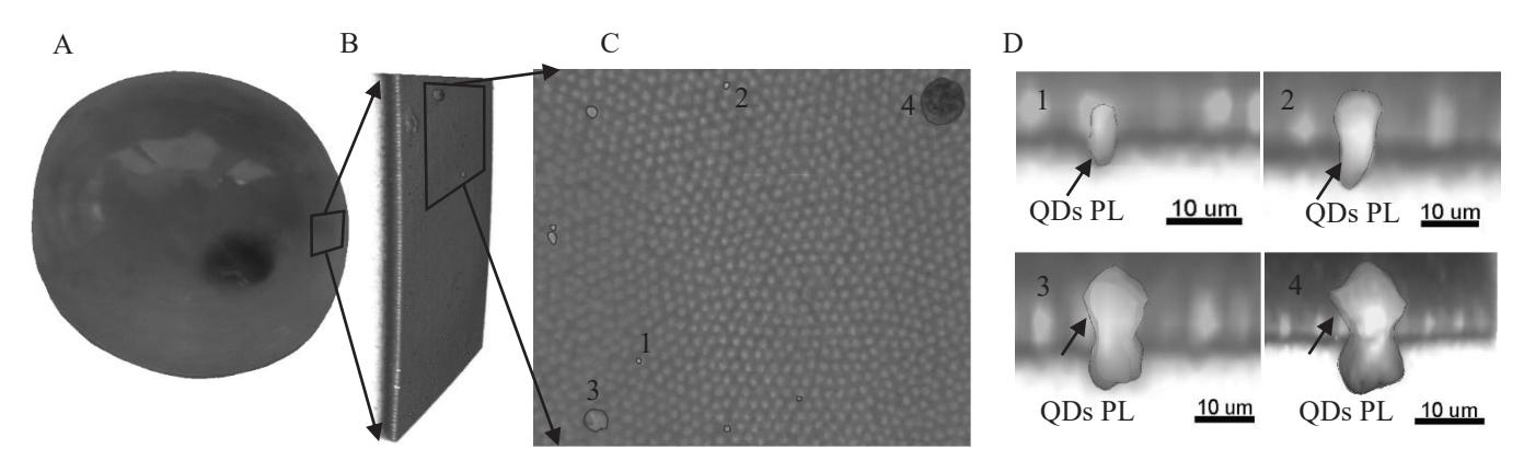
Light absorption and scattering, caused by embryo chorion tissues, allowed 3D reconstructions of external tissues with maximum thickness reaching up to 40 μm (Fig. 1. B). The autofluorescence of embryo membrane's surface is non-homogeneous (Fig. 1. C). This non-homogeneous autofluorescence can be seen as brighter spots with a diameter of 1.15 ± 0.29 μm and center to center distance of 2.27 ± 0.32 μm. TEM measurements of Oncorhynchus mykiss membrane pores has shown the same results [1]. Depending on the size of QDs conglomeration, photoluminescence of QDs can be seen directly in the area of brighter spots (Fig. 1. C – 1 and 2). As the size of QDs conglomeration increases, photoluminescence can be seen covering a wider area (Fig. 1. C – 3 and 4). A cross-section of 3D reconstruction shows a thin 3 – 4 μm external layer, composed of brighter cylindrical shaped autofluorescence structures, which coincide with the brighter spots on the surface. A part of QDs photoluminescence was observed in the area of these cylindrical shaped autofluorescence structures (Fig. 1. D – 1 and 2). As the size of QDs conglomeration increases, more dispersed QDs photoluminescence can be seen (Fig. 1. D – 3 and 4). Dispersion increases in lateral as well as vertical direction in respect to the surface of the chorion. In the case of small QDs conglomeration, QDs photoluminescence occurs up to 3 – 4 μm within the chorion. Concerning larger conglomeration, QDs photoluminescence up to 10 μm within chorion was registered.