
Recently, it has been found that electrochemical biosensors, containing gold nanoparticle (Au NP) coated ITO (indium tin oxide) electrode, exhibit high sensitivity and selectivity to detectable molecules [1,2]. That was attributed to the unique properties of Au NPs, such as large surface-to-volume ratio, high conductivity, excellent biocompatibility and low toxicity [1,3,4]. Commonly and relatively cheaply, deposition of metal nanoparticles on the glass surface is done by immersing the silanized substrate into the colloidal solution of nanoparticles [5]. However, we found that it is difficult to deposit sufficient concentration of Au NPs on the silanized ITO surface. Therefore, the approach of direct Au NP generation by nanosecond laser pulse irradiation of thin Au films [6], coated on the ITO layer, was employed. In this work, we investigated the influence of Au film thickness and laser irradiation parameters on the size distribution and area density of generated Au NPs on the ITO surface.
Firstly, seven different thicknesses of Au films were deposited on the 120 nm-thick ITO layer. Secondly, the samples were irradiated by the second harmonics (532 nm wavelength) radiation of Q-switched Nd:YVO4 laser NL220 (Ekspla), which has a pulse duration of 30 ns. When the laser radiation was absorbed in the Au film, its temperature was increased above the melting point, causing the film to rupture into nanodroplets due to the dewetting process. After the laser exposure, formed nanodroplets cooled down and solidified into Au NPs. Generated Au NPs were characterized by processing the scanning electron microscope images using the computer software ImageJ.
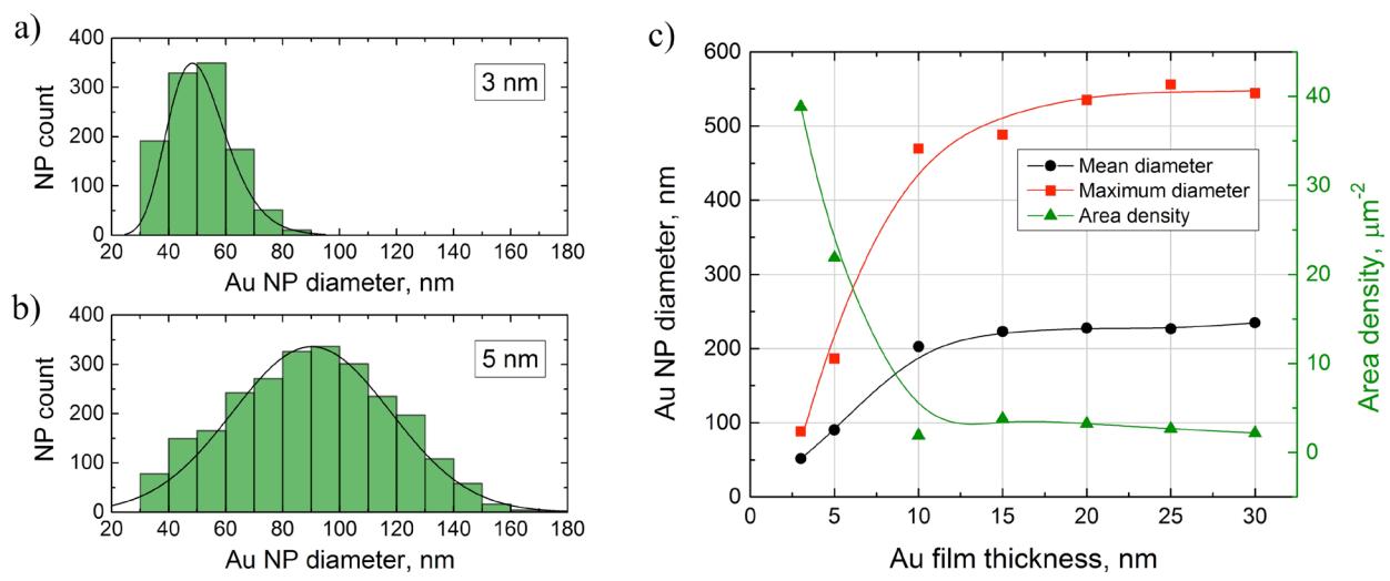
Size distribution histograms of Au NPs generated using 3 nm-thick and 5 nm-thick Au films are depicted in Figures 1a and 1b, respectively. Laser irradiation of 3 nm-thick Au film generated ~50 nm mean diameter Au NPs, whereas irradiation of 5 nm-thick Au film resulted in significantly larger size dispersion of Au NPs with a mean diameter of ~90 nm. Summarized Au NP size and area density dependence on the thickness of Au layer is shown in Figure 1c. The mean diameter reaches the value of ~220 nm and does not depend on the Au film thickness in the range of 15-30 nm. The maximum area density of generated Au NPs was ~40 μm-2 for 3 nm-thick Au film and decreased to ~3 μm-2 when generating Au NPs in 10 nm and thicker Au films.
In conclusion, it was determined how Au layer thickness and laser irradiation parameters affect the size distribution and area density of generated Au NPs. The results of this work are important for the further research to develop a highly sensitive electrochemical biosensor based on the Au NP textured ITO electrode.