
Quantum dots (QDs) - semiconductor particles of nanometre size - are widely used as photoluminescent agents in material science, biology and medicine. Enhanced photostability, size-dependent absorption and bright tuneable photoluminescence (PL) as well as wide possibilities for surface modification of QDs are among the key properties stimulating their applicability in biological environment [1]. However, the spectral properties of QDs are determined by many internal and external factors. The structure of QDs, the nature of stabilizing surface ligands [2], the surrounding environment and an irradiation can affect the stability of the PL of QDs.
In this work the spectroscopy and microscopy methods were used to estimate the light-induced changes in the PL properties of several types of QDs terminated with a carboxyl group (COOH). These core and core/shell QDs were studied in aqueous suspensions, in the presence of bovine serum albumin (BSA) and in NIH 3T3 mouse embryo fibroblast cells. The presence of BSA (Albumin, V fraction, M = 69000 g/mol, Carl Roth GmbH, Germany) molecules increased the stability and the PL quantum yield (QY) of core structured CdTe QDs and core-shell structured CdSe/ZnS QDs capped with thioglycolic (TGA) acid. However, the interaction between BSA and these core/shell QDs capped with mercaptopropionic acid (MPA) had the opposite effect. Thus, the type of terminal ligands had a strong influence on the capacity of the albumin to stabilize the PL of hydrophilic QDs through the modification of the coating layer.
The exposure to the green laser light (532 nm, 35 mW/cm2) instantly increased the PL intensity and the QY of TGA-capped core-shell QDs (Fig. 1a) irrespective of the presence or absence of BSA in solutions, but did not change the PL of MPA-capped QDs significantly. However, an instant decrease of the PL intensity and the QY, which were detected after the exposure of core QDs, mimicked the time-related changes occurring in unexposed control samples. Thus, the presence of the shell and the stability of the coating both play a role in whether their mutual effect on the PL intensity and the QY of the illuminated QDs will be suppressive or enhancing with respect to untreated samples.
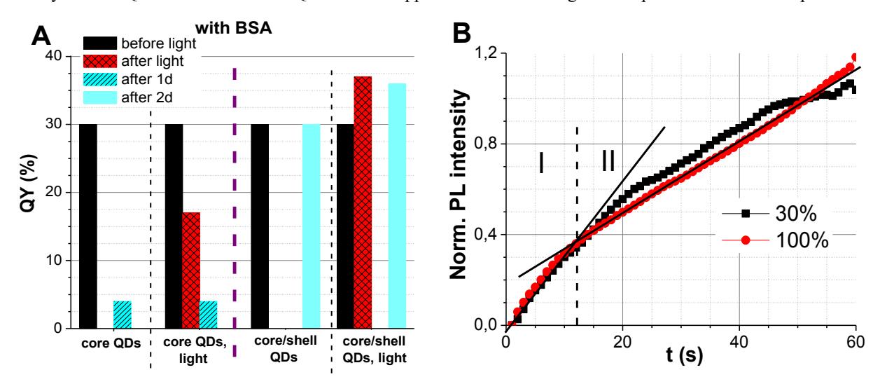
NIH 3T3 cells were incubated in a solution (c = 8 nmol/ml) with core/shell CdSe/ZnS-COOH quantum dots (Qdot 625 ITK carboxy, Invitrogen, USA) for 24 h. During subsequent exposure to the laser beam (488 nm) the PL intensity of QDs inside the cells was enhanced, and two stages were observed in the pattern of this enhancement (Fig. 1b). The increase was faster at the first stage, which lasted for initial 11-12 seconds of exposure; the increase at the second stage became slower. It has also been found that the mean PL intensity of core/shell QDs being accumulated in NIH 3T3 cells increased regardless of the excitation wavelength (488 nm or 405 nm), and during the second stage this increase of PL was directly proportional to the intensity of the excitation beam.