Time-resolved spectroscopy is particularly useful for studying properties of molecular systems since it allows to examine the processes taking place in the system and not just static system parameters. Using modern experimental setups we can study the ultrafast femto- and picosecond processes as well as the slower millisecond processes. However, to extract valuable information from the two-dimensional (time vs. wavelength) arrays of experimental data, its mathematical analysis is needed.
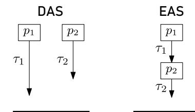
The Decay-Associated Spectra (DAS) and the Evolution-Associated Spectra (EAS) models are often incorporated [1]. By applying these models to the fluorescence data, one tries to describe the system using a number of exponentially decaying components (compartments) with their spectra and decay kinetics. The compartments used for describing the system within the DAS model are taken to be decaying independently after the initial excitation with intrinsic decay time constants $\tau_i$ (see Fig. 1). The EAS model treats the system differently, with only one component being initially excited. Its decay causes the excitation to descend sequentially to the next compartments causing them to fluoresce. After obtaining the solutions of the equations that correspond to the chosen model, we find the $\tau_i$ constants using a minimisation routine [2].
The aim of this study is to numerically simulate the time-resolved fluorescence spectra of various molecular systems and examine them using the DAS and EAS models to see whether the correct conclusions about the structure of the system can be drawn from the results of the analysis.
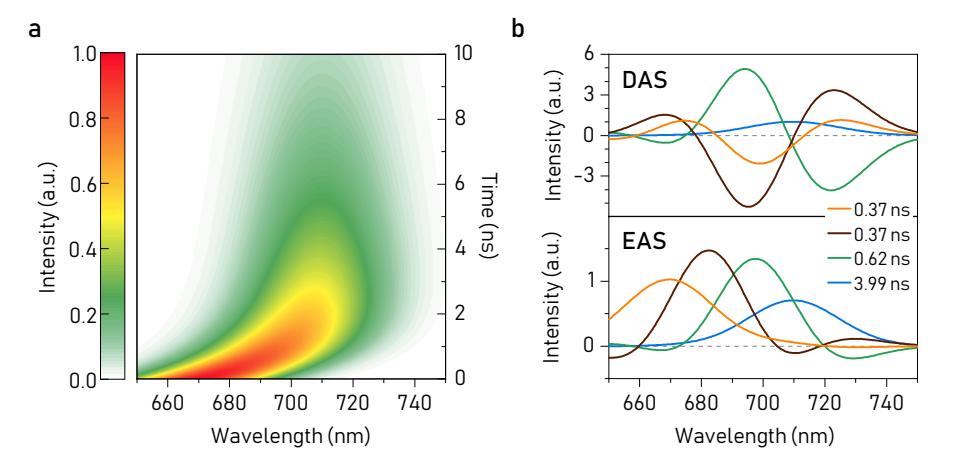
First, we discovered that the EAS model is suitable for identifying with certainty the time-dependent spectral shift [3] that might occur in the system (see Fig. 2). This finding later allowed us to distinguish two visually similar fluorescence maps produced by different systems, one of which exhibits a spectral shift. Next, we focused on a couple of systems that are composed of ensembles of monomers and dimers with randomly distributed parameters and found that the decay- and evolution-associated spectra match the spectra of the system's fluorescence sources (and thus provide a correct picture of the system) only in the simplest case of an ensemble of monomers. Finally, we studied the fluores-
cence spectra of an ensemble of the inter-connected monomers randomly arranged into a square lattice and concluded that the DAS and EAS models correctly hint at the presence of energy transfer in the system and allow us to evaluate the timescale of this process, but fail to indicate the numbers of different types of monomers found in the system.