
Every year Centers for Disease Control and Prevention and World Health Organization reports numerous cases of illnesses and even deaths, which are related with food, contaminated with pathogens [1,2]. Chlorophyllin-based photosensitization is one of the most promising innovative approaches for decontamination of fresh produce [3]. However, main disadvantage of this method is limited susceptibility of Gram-negative bacteria [4]. In order to find ways for the optimization of this technology, it is necessary to find out precise mechanism how does it destroy bacterial cell. For this reason, surface enhanced Raman scattering (SERS) was used.
The collected SERS spectra of pure E. coli, aqueous 1 mM chlorophylin solution and bacteria treated by chlorophyllin-based photosensitization are presented in the Fig. 1.
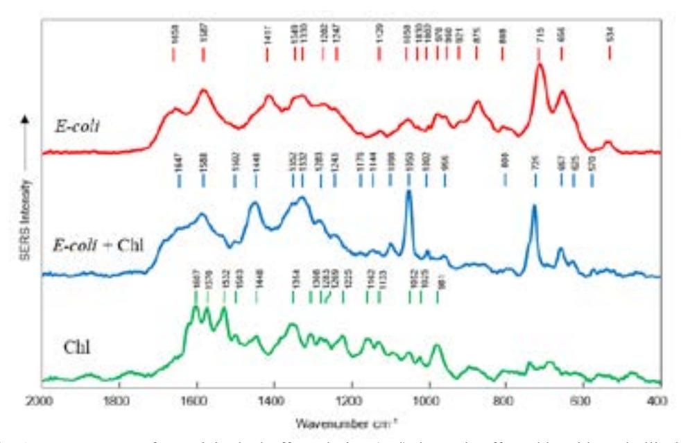
Noticeable differences in the spectra of bacteria after the treatment with chlorophyllin-based photosensitization can be seen. The major differences are the appearance of an intense spectral bands located at 1050 cm-1, 1448 cm-1 and 1098 cm-1. These bands can be attributed to the vibrations of carbohydrate fragments, $\delta$(CH2) and $\nu$(C-C) normal vibrational modes of the lipids [5]. This finding can be associated with bacteria cell membrane.
SERS spectra of these molecules are weak in the unaffected bacteria, since due to the close packing of molecules in the membrane of undamaged cells the carbohydrate molecules cannot appear in close neighbourhood with metal nanoparticles of the colloidal solution, used for Raman signal enhancement. The situation is different when the membrane is damaged - fragments of molecules from the membrane can get closer to the nanoparticles what in turn can reason strong SERS spectral bands.
These spectral features indicate that some changes related to the lipid molecules, proteins and carbohydrates are taking place due to the bacteria treatment with chlorophyllin-based photosensitization. These changes are induced by ROS [6] which lead us to believe that in fact the disruption of the integrity of the membrane is the key effect of the chlorophyllin-based photosensitization treatment.