
Silver nanoparticles (AgNPs) have remarkable antimicrobial and localized surface plasmon resonance (LSPR) properties. These AgNPs properties are reported to be dependent on the size, shape and surrounding medium. The unique properties of NPs opened broad prospects for the applications in various fields including medicine and biology [1-2]. Despite AgNPs inhibitory behavior towards few hundred species of microbes, including antibiotic resistant bacterials, AgNPs also have gained interest in gel dosimetry to enhance the dose deposited in the tumor while using low radiation as well as for better imaging purposes [3].
Most commonly used method for synthesis of silver nanoparticles is the chemical reduction of silver salts in aqueous medium (Lee-Meisel method). In certain areas of application, such as medicine, there is a need of pure AgNPs without any toxic byproducts and impurities. In such cases, mostly chemical and physical methods of AgNPs synthesis are not suitable. Then AgNPs could be produced by using irradiation of electromagnetic radiation (UV, X-ray etc.). Such techniques have several advantages over convention chemical methods, such as: i) reduction of silver ions can be carried out without excessive reducing agents or producing undesired byproducts of the reductant; ii) reducing agent is uniformly distributed in the solution; iii) radiation-induced reduction is done at ambient temperature [1-3].
In this study AgNPs in 10% gelatin medium were synthesized by UV irradiation. As silver precursor has been used silver nitrate, which concentration varied up to considerably high – 50 mM.
All materials used for AgNP synthesis were analytical grade and was used as received without any further purification. Silver nitrate (AgNO3) (Sigma Aldrich, Poland, CAS No 7761-88-8), gelatin (Sigma Aldrich, Poland, CAS No. 9000-70-8) and sodium citrate dehydrate (C5H5Na3O7·H2O) (Sigma Aldrich Reachem, Slovakia s.r.o., CAS No. 68-04-02) were used as silver precursor, main stabilizator, secondary stabilizator and reducing agent, respectively. There was prepared 6 samples, silver nitrate concentration varied from 1 mM to 50 mM, gelatin medium concentration was 10 %. Prepared samples were irradiated 60 min by UV light source (365 nm, 36 W for polymer gel curing). To eliminate the deep UV irradiation samples (<320 nm) were stored in the transparent 20 ml glass bottle. AgNPs formation, growth, and stability were analyzed by measuring UV-Vis spectrum. Spectra were measured by Ocean Optics USB4000 spectrometer at the range of 300-800 nm. AgNPs size was evaluated theoretically by applying Mie theory calculations (computer program MiePlot v4.6) [4].
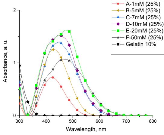
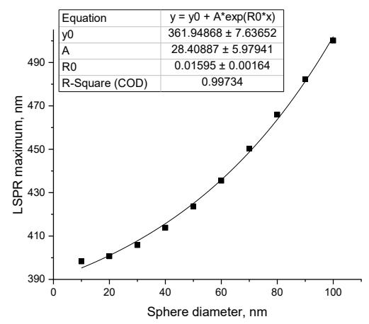
Colour of prepared silver nanoparticles solution varied from yellow to darkish brown. Synthesized samples showed very high absorption and in order to obtain visible spectrum of the LSPR samples were diluted to 25 % (fig. 1). It was measured that as silver nitrate concentration increases, peak location of the LSPR shifts towards red from 422,4 to 475,0 nm, while peak FWHM increases from 101,2 to 178,4 nm, peak height varies from 0,73 to 1,63 a. u. According to Mie calculations results, synthesized AgNPs size varied from 47,4 nm to 86,6 nm (fig. 2).