
Study proposes a new method for tow-dimensional acoustic impedance imaging for medical ultrasound B-scan images that can be performed without slicing the specimen or material as a reference. Over the last decade, there has been significant improvement in ultrasonic systems. Acoustic impedance calculation was one of the significant discoveries and uses, but no one was able to calculate it using a traditional single-element focused medical ultrasound devices. [1]
In most of optical observation of biological tissue, the specimen is sliced into several μm in thickness, and fixed on a glass substrate. On the other hand, acoustic impedance imaging can be performed without non-destructive process. The proposed method is the conversion of a B-mode image into an acoustic impedance image. The time domain reflectometry theory and transmission line model were used as reference in the calculation. Significant scatter, refraction, and attenuation were assumed not to take place during the propagation of an ultrasonic wave. Hence, they were ignored in calculations.
Two Aquaflex Ultrasound Gel Pad has been used with different characteristics were employed as the specimens to be observed. All studies have been conducted at the Department of Skin and Venereal Diseases of the Lithuanian University of Health Sciences by using Commercial DUB-USB (Taberna pro medicum, Lueneburg, Germany) single-element focused ultrasonic transducer (fundamental frequency, 22 MHz; bandwidth, 12–28 MHz, focused, focal distance – 11 mm). The radiofrequency data were analyzed in the time-frequency domain to make boundaries more noticeable. The study had been approved by regional ethics committee (Nr. P3-BE-2-25/2009).
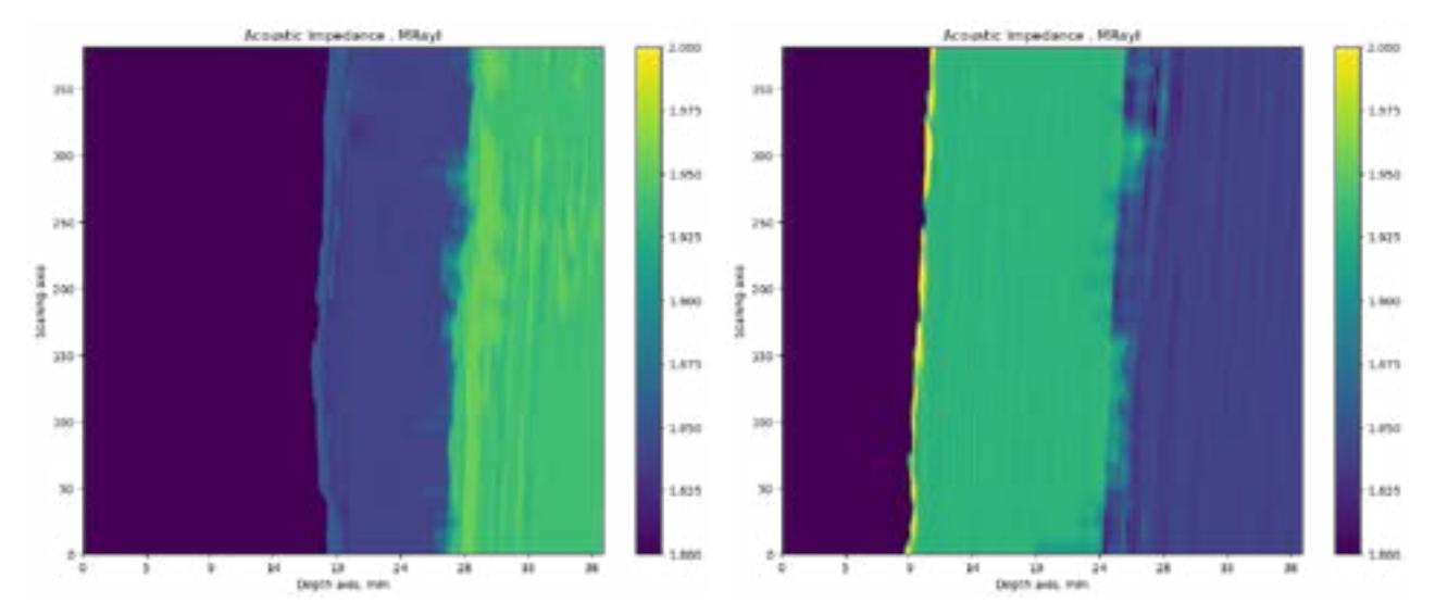
In order to calibrate the accuracy of acoustic impedance, both of phantoms with different characteristics were prepared. Their acoustic impedance was measured using density and acoustic resistance. The intensity of the reflected signal was lower with higher acoustic impedance, as the acoustic impedance of the phantoms was lower than that of the substrate. The lower signal intensity was subsequently converted into higher acoustic impedance. An example of real phantoms can be given in the figures: the first example (Fig. 1) shows an example of a transition from a lower acoustic impedance medium to a high, and a second example (Fig. 2) shows a reverse example – from a higher acoustic impedance medium goes to lower acoustic impedance medium.
The tow-dimensional acoustic impedance imaging for medical ultrasound B-scan images technique is believed to be a powerful tool for biological tissue characterization, as neither staining nor slicing is required. Some good results are obtained and hence method showed their possible applications in the study of acoustic properties of B-mode images.