
Nanotechnology is rapidly growing by producing nanoproducts and nanoparticles (NPs) that can have novel and size-related physico-chemical properties differing significantly from larger matter. This open up new horizons for application of such nanosized objects. Among all investigated nanoparticles (Au, Al, Cu, etc.), silver nanoparticles (AgNPs) have attracted increasing interest due to their unique physical, chemical and biological properties compared to their macro-scaled counterparts [1]. Nowadays AgNPs show great potential for application as antimicrobial material, in sensor chips for Raman signal enhancement, medicine, optics, etc.
The purpose of this work was to synthesize silver nanoparticles using polyol method and use these particles for signal enhancement in Raman scattering spectroscopy. The synthesis was performed in heated 1,5-Pentanediol for 8-12 min. Silver nitrate AgNO3 was used as precursor and polyvinylpyrrolidone was used as stabilizing agent to prevent particle agglomeration [2]. In the first (I) synthesis, 6 ml of AgNO3 solution was added to the synthesis medium, and 4 ml of AgNO3 – for the second (II) synthesis.
Optical properties of synthesized colloidal solutions were examined using UV-VIS spectrometry. Since the synthesized solutions are highly concentrated, the optical spectra are measured in ethanol by adding 50 μl of the test solution. Since ethanol is characterized by absorption at ~ 200 nm, the effect of ethanol could be avoided when assessing the absorption of colloidal solution, because usually silver nanoparticles have an absorption peak in a range of 350 – 800 nm depending on particle size and geometry. The measured absorption spectra and SEM micrograph of particles synthesized in II synthesis are presented in Fig. 1. and Fig. 2 respectively.
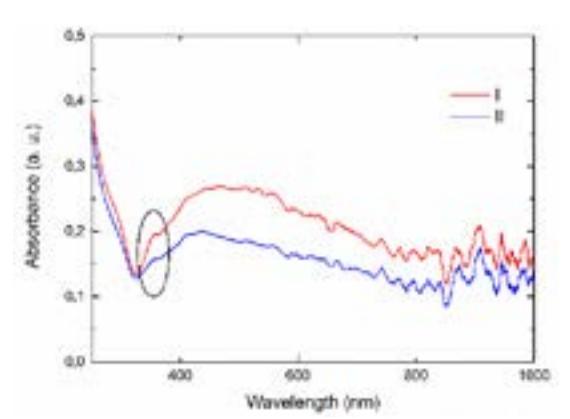
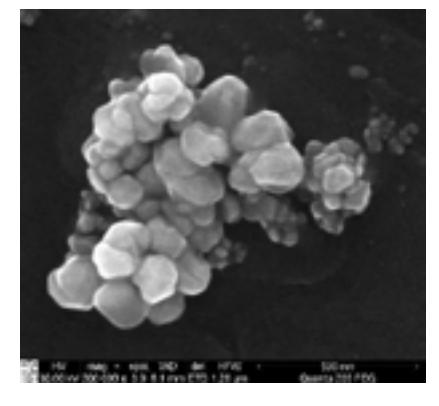
The existence of cube silver nanoparticles was confirmed by appearance of the absorbance peak around 354 nm for both synthesis (resonance due to the polarizability of the charge at the edges of the cubes [3]). Even though, the characteristic absorption peak for cube nanoparticles appeared in spectrum, microscopic analysis has shown that particle geometry varies and no predominant shape can be distinguished (Fig. 2).
Further synthesized particles were tested for SERS using 2-naphthalene thiol as analyte material. The analysis of obtained results have shown that synthesized particles can be successfully applied for SERS as the concentration of analyte molecules up to 10-4 M was detected.