Piezo materials are characterized by direct piezoelectric effect: pressure generates charges on the surface of piezoelectric materials. It will also work in reverse, the inverse piezoelectric effect causes a change in length in this type of materials when an electrical voltage is applied. Examples of piezoelectric materials are: quartz, Rochelle salt, lead titanate zirconate ceramics and bones. Piezoelectricity is good feature for bones because it can repair and heal bones. Also, bones can be remodeled with help of piezoelectricity [1].
We used Michelson interferometer with He-Ne laser in this work to measure displacement factor $\Delta h$ Eq. (1) and piezoelectric coefficient $d_{33}$ Eq. (2), due to inverse piezoelectric effect of pig rib and ox bone.
$$ \Delta h=\frac{\lambda}{4} * N \tag{1} $$$$ d_{33}=\frac{\Delta h}{U} \tag{2} $$Here $\lambda = 632.8$ nm is wavelength of laser light, N is number of light intensity changes when thickness of specimen changes, U is value of applied voltage.
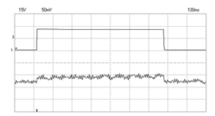
Pig rib (9 mm x 5 mm x 7 mm) and ox bone (8 mm x 5 mm x 17 mm) specimens were placed in the attachment unit. Thin mirror which moved depending on the piezo material motion was on the front surface of unit. Voltage 500V was connected to both samples and has created electric field. Signal of exiting voltage (upper curve) and signal from the photodiode (lover curve) are presented in Fig. 1. The signals of photodiode show bones thickness change. $\Delta h = 16$ nm was obtained for the pig rib, using this parameter was calculated $d_{33} = 32$ pC/N and $\Delta h = 41$ nm was obtained for ox bone, $d_{33} = 82$ pC/N.
In comparison with the first bone measured $d_{33} = 2$ pC/N (Japanese scientists: E. Fukada and I. Yasuda) [2], we received a little bit bigger coefficient. This difference can be explained that no charges leakage was in our experiments with inverse piezoelectric effect.